LectureScribe Logo
lecturescribe.io

LOCK IN VALENTINE PRICING BEFORE SALE ENDS!

Use code — New joiners get 40% OFF on annual plans

Plans will have higher prices after this sale ends

0days
:
0hours
:
0minutes
:
0seconds
Neuroscience2,500+ words5 slides

Action Potential Steps in a Neuron: Complete Guide to the Nerve Impulse

Master the action potential steps neuron science requires. Learn all 5 phases of the nerve impulse including depolarization and repolarization.

L
LectureScribeAI-Powered Study Platform

Study Infographic

Complete visual guide to the action potential steps, ion channel dynamics, and nerve impulse propagation

Download
Infographic showing the action potential steps in a neuron including depolarization, repolarization, and ion channel states

Interactive Study Short

Swipe through 5 slides about Action Potential Steps in a Neuron: Complete Guide to the Nerve Impulse

1 / 5

What Is an Action Potential?

An action potential is a rapid, transient reversal of the electrical membrane potential that travels along the axon of a neuron, serving as the fundamental unit of signaling in the nervous system. Understanding the action potential is essential for students of neuroscience, physiology, and medicine, as it underlies everything from reflexes to cognition.

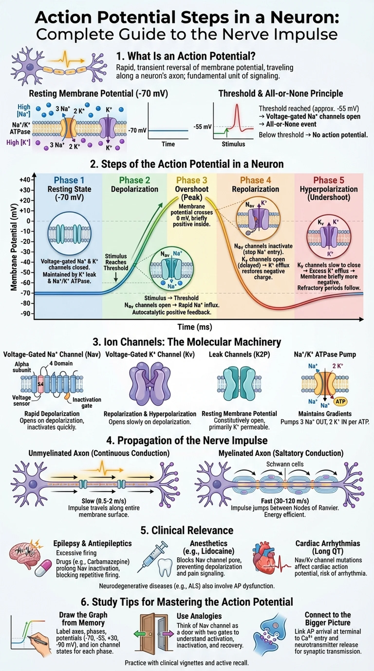
At rest, a typical neuron maintains a resting membrane potential of approximately -70 millivolts (mV), with the inside of the cell negatively charged relative to the outside. This resting potential is established primarily by the selective permeability of the membrane to potassium ions (K+) through leak channels and by the activity of the sodium-potassium ATPase pump (Na+/K+ ATPase), which actively transports three sodium ions (Na+) out of the cell for every two K+ ions pumped in. The resulting electrochemical gradients—high Na+ outside, high K+ inside—create the stored energy that drives the action potential.

When a stimulus depolarizes the membrane to a critical level called the threshold (typically around -55 mV), voltage-gated sodium channels open rapidly, allowing Na+ to flood into the cell. This triggers an all-or-none event: if threshold is reached, a full action potential fires; if it is not, no action potential occurs regardless of how close the stimulus came to threshold. This all-or-none principle means that the action potential does not vary in amplitude—information is encoded instead by the frequency of action potentials and the pattern of neurons firing.

The action potential is the basis of the nerve impulse, the electrical signal that propagates along axons to transmit information between neurons and from neurons to muscles and glands. Each nerve impulse follows an identical sequence of ion channel openings and closings, producing the characteristic spike seen on an oscilloscope or electrophysiology recording. Grasping the action potential steps in a neuron is therefore the gateway to understanding neural communication, synaptic transmission, and the pharmacology of drugs that target ion channels.

Key Terms

Action Potential

A rapid, transient reversal of membrane potential that propagates along a neuron's axon, serving as the electrical signal for neural communication.

Resting Membrane Potential

The electrical potential difference across the neuronal membrane at rest, typically about -70 mV, maintained by ion gradients and selective permeability.

Threshold

The critical level of depolarization (approximately -55 mV) that must be reached to trigger the opening of voltage-gated Na+ channels and initiate an action potential.

All-or-None Principle

The concept that an action potential either fires at full amplitude when threshold is reached or does not fire at all; there is no partial response.

Steps of the Action Potential in a Neuron

The action potential steps in a neuron can be broken down into five distinct phases: resting state, depolarization, overshoot, repolarization, and hyperpolarization (undershoot). Each phase reflects the sequential opening and closing of specific voltage-gated ion channels.

During the resting state, voltage-gated Na+ and K+ channels are closed. The membrane potential sits near -70 mV, governed by K+ leak channels and the Na+/K+ ATPase. The neuron is polarized and ready to respond to stimulation.

Depolarization begins when a stimulus—such as neurotransmitter binding at a synapse or sensory receptor activation—causes the membrane potential to become less negative. If this graded depolarization reaches threshold (around -55 mV), voltage-gated Na+ channels undergo a conformational change, opening their activation gates. Na+ rushes into the cell along its electrochemical gradient, driving the membrane potential sharply upward. This phase of depolarization is autocatalytic: the influx of Na+ depolarizes adjacent regions of the membrane, opening more Na+ channels in a positive feedback loop.

The overshoot phase occurs as the membrane potential crosses 0 mV and briefly reaches approximately +30 to +40 mV. At this point, the inside of the neuron is transiently positive relative to the outside.

Repolarization follows within about one millisecond as two events coincide. First, the inactivation gates of the voltage-gated Na+ channels close (a process called inactivation), halting Na+ influx. Second, voltage-gated K+ channels, which open more slowly than Na+ channels, reach their open state and allow K+ to flow out of the cell along its concentration gradient. This outward K+ current restores the negative charge inside the cell.

Hyperpolarization (the undershoot) occurs because the voltage-gated K+ channels are slow to close. K+ continues to leave the cell even after the membrane potential has returned to -70 mV, briefly driving it to about -80 to -90 mV. During this period, the neuron is in a relative refractory period, requiring a stronger-than-normal stimulus to fire again. The Na+/K+ ATPase then restores the original ion distributions, returning the neuron to its resting state and making it ready for the next nerve impulse.

These action potential steps repeat identically each time the neuron fires, producing the stereotyped spike waveform that is the hallmark of neural signaling.

Key Terms

Depolarization

The phase of the action potential in which the membrane potential becomes less negative (more positive) due to the influx of Na+ ions through voltage-gated channels.

Repolarization

The phase in which the membrane potential returns toward the resting value as K+ ions flow out of the cell through voltage-gated K+ channels.

Hyperpolarization

A brief overshoot below the resting membrane potential (to approximately -80 to -90 mV) caused by prolonged opening of voltage-gated K+ channels.

Inactivation Gate

A structural component of the voltage-gated Na+ channel that closes shortly after channel opening, preventing further Na+ entry and contributing to repolarization.

Refractory Period

A time interval following an action potential during which the neuron is less excitable or completely unable to fire another action potential.

Ion Channels and the Action Potential

Ion channels are the molecular machinery that executes each of the action potential steps. These transmembrane proteins form selective pores that allow specific ions to cross the lipid bilayer in response to voltage changes, ligand binding, or mechanical stimuli.

Voltage-gated sodium channels (Nav) are responsible for the rapid depolarization phase of the action potential. In mammals, nine Nav subtypes (Nav1.1 through Nav1.9) are expressed across different tissues. Each channel consists of a single large alpha subunit with four homologous domains (I–IV), each containing six transmembrane segments (S1–S6). The S4 segment in each domain acts as the voltage sensor, containing positively charged amino acids that move outward in response to depolarization, triggering channel opening. A short intracellular loop between domains III and IV serves as the inactivation gate, swinging into the pore within milliseconds to block Na+ flow—a process critical to the repolarization and refractory period phases.

Voltage-gated potassium channels (Kv) mediate repolarization and hyperpolarization. Unlike Nav channels, Kv channels are tetramers of separate alpha subunits, each with six transmembrane segments. The delayed rectifier Kv channels (e.g., Kv1, Kv2, Kv3 families) open with slower kinetics than Nav channels, which is why their peak conductance coincides with the falling phase of the action potential rather than the rising phase.

Leak channels, primarily two-pore domain potassium channels (K2P or TREK/TASK family), are constitutively open and are the dominant contributors to the resting membrane potential. Their conductance sets the baseline against which voltage-gated channel activity is measured.

The Na+/K+ ATPase is not an ion channel but an active transporter that maintains the concentration gradients essential for the action potential. By pumping three Na+ out and two K+ in per ATP hydrolyzed, it sustains the driving forces that power each nerve impulse. Importantly, the pump does not directly generate the action potential—the immediate energy source is the pre-existing ion gradients.

Pharmacological agents that target ion channels illustrate their clinical significance. Local anesthetics such as lidocaine block Nav channels, preventing depolarization and silencing pain-transmitting neurons. Tetraethylammonium (TEA) blocks Kv channels, prolonging the action potential. Understanding ion channel physiology is therefore central to neuropharmacology and the clinical management of conditions such as epilepsy, cardiac arrhythmias, and neuropathic pain.

Key Terms

Voltage-Gated Sodium Channel (Nav)

A transmembrane protein that opens in response to depolarization, allowing rapid Na+ influx that drives the rising phase of the action potential.

Voltage-Gated Potassium Channel (Kv)

A transmembrane protein that opens with delayed kinetics during depolarization, allowing K+ efflux that mediates repolarization of the neuron.

Na+/K+ ATPase

An active transporter that pumps three Na+ ions out and two K+ ions into the cell per ATP, maintaining the ionic gradients required for action potential generation.

Leak Channels

Ion channels that are constitutively open at rest, primarily permeable to K+, and are the main determinants of the resting membrane potential.

Propagation of the Nerve Impulse Along the Axon

Once generated at the axon hillock (the initial segment), the action potential must propagate along the entire length of the axon to reach synaptic terminals. The mechanism of nerve impulse propagation depends on whether the axon is myelinated or unmyelinated.

In unmyelinated axons, propagation occurs by continuous conduction. The local current generated by Na+ influx during depolarization spreads passively to adjacent regions of the membrane, depolarizing them to threshold and triggering voltage-gated Na+ channels to open in a sequential, domino-like fashion. The action potential moves in one direction because the region behind the advancing impulse is in its refractory period and cannot be re-excited. Conduction velocity in unmyelinated fibers is relatively slow, typically 0.5 to 2 meters per second, and increases with axon diameter because larger diameters reduce internal resistance to current flow.

Myelinated axons achieve much faster nerve impulse propagation through saltatory conduction. Myelin, produced by Schwann cells in the peripheral nervous system and oligodendrocytes in the central nervous system, wraps the axon in an insulating lipid sheath that prevents ion flow across the membrane. The myelin is interrupted at regular intervals by gaps called nodes of Ranvier, where voltage-gated Na+ and K+ channels are concentrated at high density. When an action potential occurs at one node, the local current jumps rapidly through the myelinated internode (due to low capacitance and high resistance of the myelin) to depolarize the next node, where a full action potential is regenerated. This saltatory (from the Latin saltare, to jump) mechanism increases conduction velocity to 30–120 meters per second in large myelinated fibers.

Saltatory conduction is not only faster but also more energy-efficient, because ion exchange occurs only at the nodes, reducing the workload of the Na+/K+ ATPase. Demyelinating diseases such as multiple sclerosis (MS) disrupt this efficient propagation. When myelin is lost, current leaks across the exposed internodal membrane, the depolarization arriving at the next node falls below threshold, and the nerve impulse fails—a phenomenon called conduction block. Clinically, this manifests as weakness, numbness, visual disturbances, and fatigue. Understanding nerve impulse propagation therefore has direct relevance to neurology and rehabilitation medicine.

The velocity of action potential propagation is a key clinical measurement. Nerve conduction studies (NCS) measure the speed and amplitude of electrically evoked nerve impulses to diagnose conditions such as carpal tunnel syndrome, Guillain-Barré syndrome, and diabetic neuropathy.

Key Terms

Saltatory Conduction

The rapid propagation of the action potential along a myelinated axon, in which the nerve impulse jumps from one node of Ranvier to the next.

Nodes of Ranvier

Gaps in the myelin sheath of a myelinated axon where voltage-gated ion channels are concentrated and action potentials are regenerated.

Myelin Sheath

An insulating lipid layer formed by glial cells (Schwann cells or oligodendrocytes) that wraps around axons to increase conduction velocity and energy efficiency.

Continuous Conduction

The slower mode of action potential propagation in unmyelinated axons, in which the impulse travels sequentially along every segment of the membrane.

Conduction Block

The failure of a nerve impulse to propagate past a demyelinated or damaged segment of an axon, as seen in diseases like multiple sclerosis.

Clinical Relevance of the Action Potential

The action potential steps are not merely academic—they are the foundation for understanding a wide spectrum of neurological and pharmacological phenomena. Disruptions at any step of the action potential can produce disease, and many therapeutics are designed to modulate specific phases of neural signaling.

Epilepsy is characterized by excessive, synchronized neuronal firing. Many antiepileptic drugs work by prolonging the inactivated state of voltage-gated Na+ channels, making it harder for neurons to fire repetitive action potentials. Carbamazepine, phenytoin, and lamotrigine all share this mechanism of use-dependent Na+ channel blockade: they bind preferentially to channels that are frequently cycling through the open and inactivated states, selectively dampening hyperactive circuits without silencing normal activity.

Local and general anesthetics exploit the action potential steps to block pain signaling or induce unconsciousness. Lidocaine and other amide local anesthetics enter the Na+ channel pore from the intracellular side and occlude it, preventing depolarization. Because small-diameter, unmyelinated C-fibers (which carry dull pain) are blocked before larger myelinated A-fibers, a differential nerve block can be achieved clinically.

Cardiac arrhythmias arise from abnormalities in the action potential of cardiac myocytes, which share the same fundamental ion channel mechanisms but have a prolonged plateau phase mediated by L-type calcium channels. Long QT syndrome, a potentially lethal arrhythmia, can result from gain-of-function mutations in Nav1.5 (the cardiac Na+ channel) or loss-of-function mutations in KCNQ1/KCNH2 (cardiac Kv channels). Understanding the action potential steps in both neurons and cardiac cells is therefore critical for pharmacologists developing antiarrhythmic drugs.

Neurodegenerative diseases also involve action potential dysfunction. In amyotrophic lateral sclerosis (ALS), motor neuron degeneration leads to progressive loss of nerve impulse transmission to skeletal muscles. Channelopathies—genetic disorders caused by mutations in ion channel genes—include conditions such as episodic ataxia, familial hemiplegic migraine, and myotonia congenita, each traceable to a specific defect in the ion channels that generate or shape the action potential.

Toxins from nature further illustrate the vulnerability of the action potential. Tetrodotoxin from puffer fish blocks Nav channels with exquisite selectivity, while batrachotoxin from poison dart frogs locks Nav channels in the open state, causing persistent depolarization. Scorpion and sea anemone toxins modify channel gating kinetics. These natural toxins have become indispensable research tools for studying the action potential steps at the molecular level.

Key Terms

Channelopathy

A disease caused by a genetic mutation in an ion channel gene, leading to abnormal channel function and disrupted electrical signaling.

Use-Dependent Block

A pharmacological mechanism in which a drug preferentially binds to and blocks ion channels that are frequently opening, selectively targeting hyperactive neurons.

Long QT Syndrome

A cardiac condition characterized by prolonged ventricular repolarization due to Na+ or K+ channel mutations, increasing the risk of potentially fatal arrhythmias.

Tetrodotoxin (TTX)

A potent neurotoxin from puffer fish that selectively blocks voltage-gated sodium channels, preventing depolarization and action potential generation.

Study Tips for Mastering the Action Potential Steps in a Neuron

The action potential is one of the most frequently tested topics in neuroscience, physiology, and medical board exams. Here are evidence-based strategies to ensure you master the action potential steps and can apply them under exam conditions.

Start by drawing the action potential graph from memory. Label the x-axis as time and the y-axis as membrane potential. Mark the resting potential (-70 mV), threshold (-55 mV), peak (+30 mV), and undershoot (-80 mV). For each phase—resting, depolarization, overshoot, repolarization, and hyperpolarization—annotate which ion channels are open, closed, or inactivated. This single diagram encapsulates the most important action potential steps and forces you to integrate channel kinetics with membrane potential changes.

Use analogies to build intuition. Think of the voltage-gated Na+ channel as a door with two gates: an activation gate (fast, opens on depolarization) and an inactivation gate (slow, closes shortly after). During rest, the activation gate is closed but the inactivation gate is open. During depolarization, both gates are briefly open, allowing Na+ flow. During inactivation, the activation gate is open but the inactivation gate has closed, blocking the pore. During recovery (return to resting potential), both gates reset to their original positions.

Connect the action potential to the bigger picture. After a nerve impulse reaches the axon terminal, it triggers Ca2+ entry through voltage-gated calcium channels, which causes synaptic vesicles to fuse with the membrane and release neurotransmitter. This links the action potential steps directly to synaptic transmission, muscle contraction, and sensory perception. Exam questions frequently test this integration.

Practice with clinical vignettes. For example: a patient with numbness and weakness is diagnosed with multiple sclerosis. Explain the pathophysiology in terms of nerve impulse propagation. Answer: demyelination causes current leak across internodes, reducing the depolarization that reaches the next node of Ranvier below threshold, resulting in conduction block and clinical symptoms.

Leverage AI-powered study tools like LectureScribe to convert your lecture recordings on action potentials into structured notes and review questions. Hearing the material, reading it, and then testing yourself creates multi-modal encoding that dramatically improves retention. Spaced repetition of these materials in the days and weeks before your exam will move the action potential steps from short-term to long-term memory.

Finally, teach the concept to someone else. Explaining the nerve impulse in your own words—from resting potential through depolarization, repolarization, and back—is one of the most effective ways to identify gaps in your understanding and solidify your knowledge.

Key Terms

Active Recall

A learning technique in which the student actively generates answers from memory rather than passively reviewing, proven to enhance long-term retention.

Spaced Repetition

A study method that schedules reviews at increasing intervals, leveraging the spacing effect to strengthen memory of material like action potential steps.

Clinical Vignette

A short clinical scenario used on medical board exams to test the application of basic science concepts, such as action potential physiology, to patient care.

Frequently Asked Questions

What are the five steps of an action potential in a neuron?

The five action potential steps are: (1) resting state at -70 mV, (2) depolarization as Na+ rushes in through voltage-gated channels, (3) overshoot to approximately +30 mV, (4) repolarization as K+ flows out, and (5) hyperpolarization (undershoot) below resting potential before returning to baseline.

What causes depolarization during the action potential?

Depolarization is caused by the rapid opening of voltage-gated sodium channels when the membrane reaches threshold (about -55 mV). The influx of positively charged Na+ ions drives the membrane potential from -70 mV toward +30 mV in a positive feedback loop.

What is the difference between absolute and relative refractory periods?

During the absolute refractory period, voltage-gated Na+ channels are inactivated and no stimulus can trigger another action potential. During the relative refractory period, some Na+ channels have recovered but K+ channels are still open, so a stronger-than-normal stimulus is required to fire another nerve impulse.

How does saltatory conduction speed up the nerve impulse?

In myelinated axons, the myelin sheath insulates the axon between nodes of Ranvier. The action potential jumps from node to node rather than traveling continuously along the membrane, increasing conduction velocity from about 1 m/s in unmyelinated fibers to up to 120 m/s in large myelinated fibers.

What happens to the action potential in multiple sclerosis?

In multiple sclerosis, the immune system damages the myelin sheath surrounding axons. Without myelin, current leaks across the exposed membrane during saltatory conduction, and the depolarization reaching the next node of Ranvier may fall below threshold, causing conduction block and neurological symptoms.

Why is the action potential described as all-or-none?

The action potential is all-or-none because once the threshold is reached, the full action potential fires at maximum amplitude regardless of stimulus strength. If threshold is not reached, no action potential occurs. Information is encoded by firing frequency, not by the size of individual action potentials.

How do local anesthetics like lidocaine affect the action potential?

Local anesthetics block voltage-gated sodium channels from the intracellular side, preventing Na+ influx and thus blocking depolarization. Without depolarization, no action potential is generated in the affected nerve fibers, which eliminates pain sensation in the targeted area.

Study Neuroscience Smarter

Upload your lecture notes and get AI-generated flashcards, quizzes, infographics, and study guides tailored to your curriculum.

Try LectureScribe Free

Create Study Materials

Transform your Neuroscience lectures into flashcards, quizzes, and visual study guides with AI.

  • AI-generated flashcards & quizzes
  • Visual infographics from notes
  • Interactive study shorts
Start Studying Free

Quick Facts

SubjectNeuroscience
Word Count2,500+
Slides5
InfographicIncluded
AuthorLectureScribe

Master Neuroscience with LectureScribe

Upload your lecture notes and get AI-powered flashcards, quizzes, infographics, and study guides in minutes.