LectureScribe Logo
lecturescribe.io

LOCK IN VALENTINE PRICING BEFORE SALE ENDS!

Use code — New joiners get 40% OFF on annual plans

Plans will have higher prices after this sale ends

0days
:
0hours
:
0minutes
:
0seconds
Anatomy2,500+ words5 slides
💪

Brachial Plexus Anatomy: Complete Guide to Nerves and Upper Limb Innervation

Master brachial plexus anatomy with our complete guide covering roots, trunks, divisions, cords, branches, and upper limb innervation for medical students.

L
LectureScribeAI-Powered Study Platform

Study Infographic

Complete visual overview of brachial plexus anatomy from roots (C5-T1) to terminal branches

Download
Brachial plexus anatomy diagram showing roots trunks divisions cords and branches with labeled nerves

Interactive Study Short

Swipe through 5 slides about Brachial Plexus Anatomy: Complete Guide to Nerves and Upper Limb Innervation

1 / 5

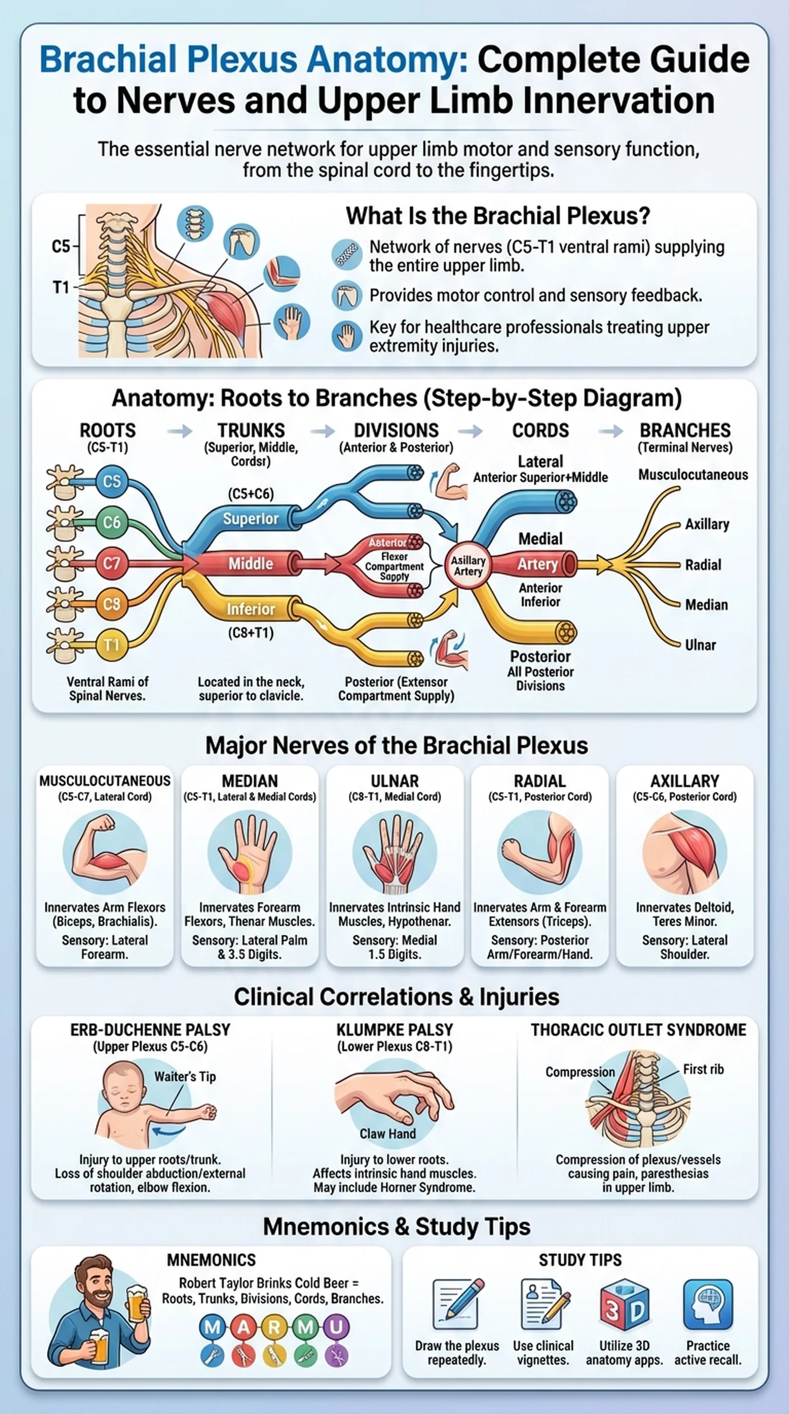
What Is the Brachial Plexus?

The brachial plexus is one of the most clinically significant nerve networks in the human body. It is a complex arrangement of nerve fibers that originates from the spinal cord and provides motor and sensory innervation to the entire upper limb, including the shoulder, arm, forearm, and hand. Understanding brachial plexus anatomy is essential for medical students, physical therapists, and any healthcare professional who treats injuries of the upper extremity.

The brachial plexus is formed by the ventral rami of spinal nerves C5, C6, C7, C8, and T1. These nerve roots emerge from the intervertebral foramina of the cervical and upper thoracic spine and converge in the posterior triangle of the neck before passing into the axilla. The network undergoes a series of merging and branching events that ultimately produce the named peripheral nerves responsible for upper limb innervation. This intricate organization allows the nervous system to distribute fibers from multiple spinal levels to individual muscles and skin regions, providing redundancy and precise motor control.

Brachial plexus anatomy is a cornerstone of gross anatomy courses and appears frequently on examinations such as the USMLE, COMLEX, and NBME shelf exams. Clinicians encounter brachial plexus injuries in trauma settings, obstetric complications, and surgical procedures involving the neck and shoulder. A firm grasp of the plexus structure enables accurate localization of nerve lesions based on a patient's motor and sensory deficits. For these reasons, dedicating focused study time to the brachial plexus pays dividends throughout a medical career.

In the sections that follow, we will walk through each organizational level of the brachial plexus, identify the major brachial plexus nerves that arise from it, discuss clinical correlations and common injuries, and share proven mnemonics and study strategies to help you retain this material efficiently.

Key Terms

Brachial Plexus

A network of nerves formed by the ventral rami of C5-T1 that provides motor and sensory innervation to the upper limb.

Ventral Ramus

The anterior division of a spinal nerve that supplies the limbs and the anterolateral trunk.

Upper Limb Innervation

The distribution of motor and sensory nerve fibers to the shoulder, arm, forearm, and hand via the brachial plexus.

Posterior Triangle of the Neck

An anatomical region bounded by the sternocleidomastoid, trapezius, and clavicle through which the brachial plexus passes.

Spinal Nerve Roots

Bundles of nerve fibers emerging from the spinal cord that combine to form the brachial plexus (C5-T1).

Anatomy of the Brachial Plexus: Roots, Trunks, Divisions, Cords, and Branches

The anatomy of the brachial plexus is organized into five structural levels: roots, trunks, divisions, cords, and branches. Each level represents a stage at which nerve fibers regroup before ultimately forming the terminal nerves of the upper limb. Learning these levels in order is the key to mastering brachial plexus anatomy.

The roots of the brachial plexus are the ventral rami of C5, C6, C7, C8, and T1. They emerge between the anterior and middle scalene muscles in the neck. At this level, several nerves branch off directly, including the dorsal scapular nerve (C5) and the long thoracic nerve (C5, C6, C7). These are sometimes called supraclavicular branches because they arise before the plexus crosses the clavicle.

The roots then merge to form three trunks. The superior trunk is formed by the union of C5 and C6. The middle trunk is a continuation of C7 alone. The inferior trunk is formed by C8 and T1. The trunks are located in the posterior triangle of the neck, superior to the clavicle. The suprascapular nerve and the nerve to the subclavius arise from the superior trunk at this level.

Each trunk divides into an anterior and a posterior division, producing six divisions in total. The anterior divisions generally supply flexor compartments of the upper limb, while the posterior divisions supply extensor compartments. This functional separation is a fundamental organizing principle of upper limb innervation.

The divisions regroup to form three cords, named by their relationship to the axillary artery. The lateral cord is formed by the anterior divisions of the superior and middle trunks (C5, C6, C7). The medial cord is a continuation of the anterior division of the inferior trunk (C8, T1). The posterior cord is formed by all three posterior divisions (C5-T1). The cords give off several important branches before terminating as the major brachial plexus nerves.

From the cords emerge the five terminal branches: the musculocutaneous nerve, the median nerve, the ulnar nerve, the radial nerve, and the axillary nerve. Additional branches from the cords include the medial pectoral nerve, lateral pectoral nerve, subscapular nerves, and thoracodorsal nerve. Understanding this hierarchical structure is essential for predicting which muscles and skin areas will be affected by injuries at different levels of the plexus.

Key Terms

Trunks

Three bundles (superior, middle, inferior) formed by the merging of the C5-T1 roots of the brachial plexus.

Divisions

Anterior and posterior branches of each trunk that separate fibers destined for flexor and extensor compartments.

Cords

Three bundles (lateral, medial, posterior) named by their position relative to the axillary artery, formed by regrouping of divisions.

Lateral Cord

Formed by the anterior divisions of the superior and middle trunks; gives rise to the musculocutaneous nerve and the lateral root of the median nerve.

Posterior Cord

Formed by all three posterior divisions; gives rise to the axillary and radial nerves.

Major Nerves of the Brachial Plexus

The brachial plexus nerves that emerge as terminal branches are responsible for the motor and sensory functions of the entire upper limb. Each nerve follows a distinct anatomical course and innervates specific muscles and cutaneous regions. A systematic understanding of these nerves is critical for clinical diagnosis.

The musculocutaneous nerve (C5, C6, C7) arises from the lateral cord and pierces the coracobrachialis muscle. It innervates the flexors of the arm, including the biceps brachii and brachialis. After passing through these muscles, it continues as the lateral cutaneous nerve of the forearm, providing sensation to the lateral forearm. Injury to this nerve results in weakness of elbow flexion and loss of the biceps reflex.

The median nerve (C5-T1) is formed by contributions from both the lateral and medial cords. It descends along the medial aspect of the arm, passes through the cubital fossa, and enters the forearm between the two heads of the pronator teres. The median nerve innervates most of the forearm flexors and the thenar muscles of the hand. It also provides sensory innervation to the palmar surface of the lateral three and a half digits. Carpal tunnel syndrome, one of the most common compression neuropathies, involves the median nerve at the wrist.

The ulnar nerve (C8, T1) arises from the medial cord, passes posterior to the medial epicondyle of the humerus at the elbow, and enters the hand through Guyon's canal. It innervates the intrinsic muscles of the hand, including the interossei and the medial two lumbricals, as well as the hypothenar muscles. Sensory distribution covers the medial one and a half digits. The ulnar nerve is vulnerable at the elbow, where it can be compressed or stretched, producing numbness and tingling in the ring and little fingers.

The radial nerve (C5-T1) is the largest terminal branch of the posterior cord. It spirals around the humerus in the radial groove, supplying the triceps and the extensors of the forearm and wrist. Sensory branches cover the posterior arm, forearm, and the dorsal aspect of the lateral three and a half digits. Radial nerve injury in the spiral groove, often associated with humeral shaft fractures, results in wrist drop due to paralysis of the wrist extensors.

The axillary nerve (C5, C6) branches from the posterior cord and passes through the quadrangular space. It innervates the deltoid and teres minor muscles and provides sensation to the regimental badge area of the lateral shoulder. Anterior shoulder dislocations frequently injure the axillary nerve, leading to weakness in arm abduction beyond fifteen degrees.

Key Terms

Musculocutaneous Nerve

A terminal branch of the lateral cord (C5-C7) that innervates the flexors of the arm and provides sensation to the lateral forearm.

Median Nerve

A terminal branch formed by the lateral and medial cords (C5-T1) that innervates forearm flexors and thenar muscles, with sensation to the lateral palm.

Ulnar Nerve

A terminal branch of the medial cord (C8-T1) that innervates most intrinsic hand muscles and provides sensation to the medial one and a half digits.

Radial Nerve

The largest branch of the posterior cord (C5-T1) that innervates the extensors of the arm and forearm.

Axillary Nerve

A branch of the posterior cord (C5-C6) that innervates the deltoid and teres minor and supplies sensation to the lateral shoulder.

Clinical Correlations and Brachial Plexus Injuries

Brachial plexus injuries are encountered across multiple medical specialties, including orthopedics, neurology, neurosurgery, and obstetrics. These injuries range from mild traction neuropraxia to complete avulsion of nerve roots from the spinal cord. Understanding the anatomy of the brachial plexus allows clinicians to localize the injury and predict functional deficits.

Erb-Duchenne palsy, also known as upper brachial plexus injury, involves the C5 and C6 nerve roots or the superior trunk. It is the most common form of obstetric brachial plexus injury and occurs when excessive lateral flexion of the neck during delivery stretches the upper roots. The affected infant presents with the arm adducted, internally rotated, and extended at the elbow, a posture known as the waiter's tip position. The deltoid, biceps, brachialis, and supraspinatus are primarily affected, resulting in loss of arm abduction, external rotation, and elbow flexion.

Klumpke palsy is a lower brachial plexus injury involving C8 and T1. It may occur during a difficult delivery when the arm is pulled overhead or in adults who grab an object while falling. The intrinsic muscles of the hand are affected, producing a claw hand deformity. If the T1 root is involved proximal to the sympathetic chain, an ipsilateral Horner syndrome (ptosis, miosis, anhidrosis) may also be present.

Traumatic brachial plexus injuries in adults often result from motorcycle accidents, falls, or contact sports. High-velocity traction injuries can avulse nerve roots directly from the spinal cord, a devastating injury that is not amenable to direct nerve repair. Findings such as a positive Tinel sign, electromyographic changes, and MRI evidence of pseudomeningocele formation help determine the level and severity of injury.

Thoracic outlet syndrome is a clinical condition in which the brachial plexus or subclavian vessels are compressed as they pass through the thoracic outlet between the scalene muscles and the first rib. Patients may experience upper limb pain, paresthesias, and weakness, particularly with overhead activities. Treatment ranges from physical therapy and postural correction to surgical decompression in refractory cases. Accurate knowledge of brachial plexus anatomy is indispensable for diagnosing and managing all of these conditions.

Key Terms

Erb-Duchenne Palsy

An upper brachial plexus injury (C5-C6) presenting with the waiter's tip position due to loss of shoulder abduction, external rotation, and elbow flexion.

Klumpke Palsy

A lower brachial plexus injury (C8-T1) resulting in claw hand deformity and possible Horner syndrome.

Thoracic Outlet Syndrome

Compression of the brachial plexus or subclavian vessels between the scalene muscles and the first rib, causing upper limb pain and paresthesias.

Neuropraxia

A mild nerve injury involving temporary loss of conduction without structural damage, with full recovery expected.

Horner Syndrome

A clinical triad of ptosis, miosis, and anhidrosis resulting from disruption of sympathetic fibers, sometimes associated with T1 root avulsion.

Mnemonics for the Brachial Plexus

The complexity of brachial plexus anatomy makes it one of the most mnemonic-dependent topics in medical education. Well-crafted mnemonics can dramatically accelerate memorization and improve recall during high-stakes examinations. Below are some of the most popular and effective memory aids used by students worldwide.

The most widely used mnemonic for the organizational levels of the brachial plexus is "Robert Taylor Drinks Cold Beer." Each word corresponds to a structural level: Roots, Trunks, Divisions, Cords, and Branches. This simple phrase provides an immediate framework for recalling the five levels in order, which is the foundation of understanding the entire plexus.

To remember the nerve roots that form the brachial plexus, students often use "C5, C6, C7, C8, T1 — five roots make the plexus fun." While simple, this rhyme reinforces the specific spinal levels involved and helps prevent confusion with other nerve plexuses such as the cervical or lumbar plexus.

For the terminal branches, the mnemonic "My Aunt Roberta Urinates Mustard" corresponds to Musculocutaneous, Axillary, Radial, Ulnar, and Median nerves. Some students prefer the variant "MARMU" as an acronym. The key is choosing a mnemonic that creates a vivid mental image, as research on memory encoding consistently shows that emotional or absurd associations are retained far better than abstract lists.

The relationships between cords and terminal branches can be recalled with the following associations. The lateral cord gives rise to the lateral pectoral nerve, the musculocutaneous nerve, and the lateral contribution to the median nerve. Think "LML" — Lateral pectoral, Musculocutaneous, Lateral root of median. The medial cord yields the medial pectoral nerve, medial cutaneous nerves of the arm and forearm, the ulnar nerve, and the medial contribution to the median nerve. The posterior cord gives rise to the subscapular nerves, the thoracodorsal nerve, the axillary nerve, and the radial nerve. The mnemonic "STAR" helps: Subscapular, Thoracodorsal, Axillary, Radial.

For remembering brachial plexus nerves and their root values, use "C5-C6 pick up sticks" for the musculocutaneous nerve and "C5-C6-C7-C8-T1" for the median and radial nerves, which draw from the broadest range of roots. Combining multiple mnemonics into a study sheet and reviewing it daily using spaced repetition will lock this material into long-term memory.

Key Terms

Robert Taylor Drinks Cold Beer

A mnemonic for the five structural levels of the brachial plexus: Roots, Trunks, Divisions, Cords, Branches.

STAR Mnemonic

A mnemonic for branches of the posterior cord: Subscapular, Thoracodorsal, Axillary, Radial.

Spaced Repetition

A learning technique that involves reviewing material at increasing intervals to strengthen long-term retention.

Terminal Branches

The five final nerves of the brachial plexus: musculocutaneous, axillary, radial, ulnar, and median.

Study Tips for Mastering Brachial Plexus Anatomy

Successfully learning brachial plexus anatomy requires a multi-modal study approach that goes beyond passive reading. The plexus is a three-dimensional structure, and students who engage with it through drawing, visualization, and active recall consistently outperform those who rely solely on textbook diagrams.

One of the most effective strategies is to draw the brachial plexus from memory repeatedly. Start by sketching the five roots (C5-T1) on the left side of a blank page. Then draw lines merging into three trunks, splitting into six divisions, reforming into three cords, and finally branching into the five terminal nerves. Label every structure as you go. Doing this exercise daily for a week will build a strong mental map of the entire plexus. Many students use color-coding, assigning different colors to each cord and its branches, which adds a visual dimension that enhances encoding.

Clinical case-based learning is another powerful approach. Rather than memorizing isolated facts, work through clinical vignettes that require you to identify the injured nerve based on motor and sensory findings. For example, if a patient presents with wrist drop after a humeral shaft fracture, you should be able to trace the deficit to the radial nerve in the spiral groove. These kinds of applied questions are exactly what you will encounter on board examinations and in clinical rotations.

Cadaveric dissection or prosection review, when available, offers unmatched spatial understanding of upper limb innervation. If you do not have access to a cadaver lab, high-quality anatomy atlases and 3D anatomy apps can serve as effective substitutes. Rotating a digital model of the brachial plexus and examining it from different angles helps solidify the spatial relationships between nerves, vessels, and muscles.

Group study sessions focused on the brachial plexus can be highly productive. Quiz each other on nerve root values, motor functions, sensory distributions, and clinical scenarios. Teaching the material to a peer is one of the most effective ways to identify gaps in your own understanding. Finally, integrate your brachial plexus study with related topics such as the cranial nerves and the action potential in neurons. Seeing the connections between nervous system topics builds a cohesive framework that supports long-term retention and clinical reasoning.

LectureScribe's AI-generated study materials, including slides, infographics, and practice questions, can supplement your brachial plexus study by providing spaced repetition prompts and visual summaries that reinforce the core concepts covered in this guide.

Key Terms

Active Recall

A study technique that involves actively retrieving information from memory rather than passively reviewing notes.

Clinical Vignette

A short clinical scenario used in medical education to test the application of basic science knowledge to patient care.

Cadaveric Dissection

The systematic study of human anatomy by dissecting a preserved human body, considered the gold standard for learning structural relationships.

Color-Coding

A visual study strategy that assigns different colors to related anatomical structures to enhance memory encoding.

Frequently Asked Questions

What are the five structural levels of the brachial plexus anatomy?

The brachial plexus is organized into roots, trunks, divisions, cords, and branches. The roots (C5-T1) merge into three trunks, split into six divisions, reform into three cords, and finally give rise to the terminal branches that innervate the upper limb.

What spinal nerve roots form the brachial plexus?

The brachial plexus is formed by the ventral rami of spinal nerves C5, C6, C7, C8, and T1. These roots emerge from the cervical and upper thoracic spine and merge to form the trunks of the plexus.

What are the major brachial plexus nerves?

The five terminal brachial plexus nerves are the musculocutaneous nerve, median nerve, ulnar nerve, radial nerve, and axillary nerve. Each provides specific motor and sensory innervation to regions of the upper limb.

What is upper limb innervation?

Upper limb innervation refers to the supply of motor and sensory nerve fibers to the shoulder, arm, forearm, and hand. The brachial plexus is the primary source of upper limb innervation, distributing fibers from spinal levels C5-T1.

What is Erb-Duchenne palsy?

Erb-Duchenne palsy is an upper brachial plexus injury affecting C5-C6 that results in the waiter's tip position. It commonly occurs during difficult deliveries when the neck is laterally flexed, stretching the superior trunk.

What is the best mnemonic for the brachial plexus?

The most popular mnemonic is 'Robert Taylor Drinks Cold Beer,' which represents Roots, Trunks, Divisions, Cords, and Branches. For the posterior cord branches, 'STAR' (Subscapular, Thoracodorsal, Axillary, Radial) is widely used.

How does the brachial plexus relate to thoracic outlet syndrome?

In thoracic outlet syndrome, the brachial plexus or subclavian vessels are compressed as they pass between the scalene muscles and the first rib. This compression can cause pain, paresthesias, and weakness in the upper limb.

Study Anatomy Smarter

Upload your lecture notes and get AI-generated flashcards, quizzes, infographics, and study guides tailored to your curriculum.

Try LectureScribe Free

Create Study Materials

Transform your Anatomy lectures into flashcards, quizzes, and visual study guides with AI.

  • AI-generated flashcards & quizzes
  • Visual infographics from notes
  • Interactive study shorts
Start Studying Free

Quick Facts

SubjectAnatomy
Word Count2,500+
Slides5
InfographicIncluded
AuthorLectureScribe

Master Anatomy with LectureScribe

Upload your lecture notes and get AI-powered flashcards, quizzes, infographics, and study guides in minutes.