LectureScribe Logo
lecturescribe.io

LOCK IN VALENTINE PRICING BEFORE SALE ENDS!

Use code — New joiners get 40% OFF on annual plans

Plans will have higher prices after this sale ends

0days
:
0hours
:
0minutes
:
0seconds
Anatomy2,500+ words5 slides
🧠

Cranial Nerves Mnemonic: Learn All 12 Cranial Nerves and Their Functions

Memorize the 12 cranial nerves with proven mnemonics. Learn cranial nerve functions, sensory vs motor classification, and clinical testing methods.

L
LectureScribeAI-Powered Study Platform

Study Infographic

Visual reference chart of all 12 cranial nerves, their functions, sensory/motor classification, and popular mnemonic devices

Download
Infographic showing the 12 cranial nerves with their names, numbers, functions, and a cranial nerves mnemonic

Interactive Study Short

Swipe through 5 slides about Cranial Nerves Mnemonic: Learn All 12 Cranial Nerves and Their Functions

1 / 5

What Are the Cranial Nerves?

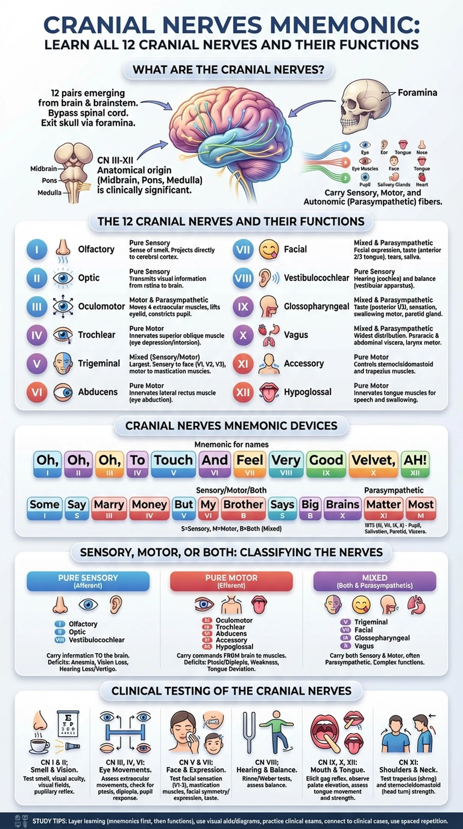
The cranial nerves are twelve pairs of nerves that emerge directly from the brain and brainstem, bypassing the spinal cord to innervate structures primarily in the head and neck. Unlike spinal nerves, which exit through the vertebral column, the cranial nerves pass through foramina (openings) in the skull to reach their target tissues. A thorough understanding of the 12 cranial nerves is essential for students of anatomy, neuroscience, and clinical medicine.

Each cranial nerve is designated by a Roman numeral (I through XII) based on the order in which it exits the brain, from anterior to posterior. Cranial nerve I (olfactory) and cranial nerve II (optic) are unique in that they emerge from the cerebrum and diencephalon, respectively, rather than the brainstem. Cranial nerves III through XII arise from the midbrain, pons, or medulla oblongata. This anatomical origin has clinical significance: a lesion in the midbrain may affect CN III, while a medullary stroke may impair CN IX, X, XI, or XII.

The cranial nerves carry sensory information (such as vision, hearing, taste, and smell), motor commands (to muscles of the eyes, face, tongue, and pharynx), and autonomic (parasympathetic) fibers that regulate pupil constriction, salivation, lacrimation, and visceral organ function. Some cranial nerves are purely sensory (I, II, VIII), some are purely motor (III, IV, VI, XI, XII), and others are mixed, carrying both sensory and motor fibers (V, VII, IX, X). This sensory-motor classification is itself a popular exam topic and the subject of its own cranial nerves mnemonic.

Learning the cranial nerves is a rite of passage in medical and health sciences education. The volume of detail—names, numbers, functions, pathways, foramina, and clinical correlations—can feel overwhelming. That is precisely why cranial nerves mnemonic devices have been used by generations of students to organize and retain this information. With the right mnemonics and a structured study approach, you can master the 12 cranial nerves efficiently and apply your knowledge in clinical settings.

Key Terms

Cranial Nerves

Twelve pairs of nerves that arise directly from the brain and brainstem, innervating structures of the head, neck, and (in the case of CN X) thoracic and abdominal viscera.

Foramen

An opening or passage in a bone through which a cranial nerve, blood vessel, or other structure passes; plural: foramina.

Brainstem

The posterior part of the brain consisting of the midbrain, pons, and medulla oblongata, from which cranial nerves III–XII originate.

Parasympathetic Fibers

Autonomic nerve fibers carried by certain cranial nerves (III, VII, IX, X) that regulate involuntary functions such as pupil constriction, glandular secretion, and heart rate.

The 12 Cranial Nerves and Their Functions

A systematic review of each of the 12 cranial nerves and their functions provides the foundation for applying any cranial nerves mnemonic. Here is a concise overview of each nerve.

CN I, the Olfactory nerve, is a purely sensory nerve that carries the sense of smell from the nasal mucosa to the olfactory bulb. It is the only cranial nerve that projects directly to the cerebral cortex without first relaying through the thalamus.

CN II, the Optic nerve, transmits visual information from the retina to the lateral geniculate nucleus of the thalamus and then to the primary visual cortex. It is technically a tract of the central nervous system, myelinated by oligodendrocytes rather than Schwann cells.

CN III, the Oculomotor nerve, innervates four of the six extraocular muscles (superior rectus, inferior rectus, medial rectus, inferior oblique) and the levator palpebrae superioris. It also carries parasympathetic fibers to the pupillary sphincter (causing pupil constriction) and the ciliary muscle (enabling accommodation).

CN IV, the Trochlear nerve, innervates the superior oblique muscle, which depresses and intorts the eye. It is the only cranial nerve that exits from the dorsal aspect of the brainstem and has the longest intracranial course.

CN V, the Trigeminal nerve, is the largest cranial nerve. It has three divisions: V1 (ophthalmic), V2 (maxillary), and V3 (mandibular). It provides sensory innervation to the face and motor innervation to the muscles of mastication.

CN VI, the Abducens nerve, innervates the lateral rectus muscle, which abducts the eye. A CN VI palsy produces medial deviation (esotropia) of the affected eye.

CN VII, the Facial nerve, controls the muscles of facial expression, carries taste from the anterior two-thirds of the tongue, and provides parasympathetic innervation to the submandibular, sublingual, and lacrimal glands.

CN VIII, the Vestibulocochlear nerve, carries auditory information from the cochlea and balance information from the vestibular apparatus to the brainstem.

CN IX, the Glossopharyngeal nerve, provides taste and sensation from the posterior one-third of the tongue, motor innervation to the stylopharyngeus muscle, and parasympathetic innervation to the parotid gland.

CN X, the Vagus nerve, is the longest cranial nerve, extending into the thorax and abdomen. It provides motor innervation to the pharynx and larynx, parasympathetic innervation to thoracic and abdominal viscera, and sensory innervation from the larynx, viscera, and a small area of the ear.

CN XI, the Accessory nerve (spinal accessory), innervates the sternocleidomastoid and trapezius muscles, controlling head rotation and shoulder elevation.

CN XII, the Hypoglossal nerve, innervates the intrinsic and most extrinsic muscles of the tongue, controlling tongue movement essential for speech and swallowing.

Knowing the cranial nerve functions in this level of detail is the prerequisite for effective use of any cranial nerves mnemonic device.

Key Terms

Olfactory Nerve (CN I)

The first cranial nerve; a purely sensory nerve responsible for the sense of smell, projecting from the nasal epithelium to the olfactory bulb.

Vagus Nerve (CN X)

The tenth cranial nerve; a mixed nerve with the widest distribution of any cranial nerve, providing parasympathetic innervation to thoracic and abdominal organs.

Trigeminal Nerve (CN V)

The fifth and largest cranial nerve; provides sensory innervation to the face (three divisions) and motor innervation to the muscles of mastication.

Facial Nerve (CN VII)

The seventh cranial nerve; controls muscles of facial expression, carries taste from the anterior two-thirds of the tongue, and provides parasympathetic innervation to salivary and lacrimal glands.

Hypoglossal Nerve (CN XII)

The twelfth cranial nerve; a purely motor nerve that innervates the tongue muscles, essential for speech, mastication, and swallowing.

Cranial Nerves Mnemonic Devices

Mnemonic devices are among the most effective tools for memorizing the 12 cranial nerves in order. A good cranial nerves mnemonic encodes the names (or their first letters) into a memorable phrase, reducing the cognitive load of rote memorization. Here are the most widely used and classroom-tested mnemonics.

The classic cranial nerves mnemonic for the nerve names uses the first letter of each nerve in order (Olfactory, Optic, Oculomotor, Trochlear, Trigeminal, Abducens, Facial, Vestibulocochlear, Glossopharyngeal, Vagus, Accessory, Hypoglossal): "Oh, Oh, Oh, To Touch And Feel Very Good Velvet, AH!" Each word corresponds to one cranial nerve: Oh = Olfactory, Oh = Optic, Oh = Oculomotor, To = Trochlear, Touch = Trigeminal, And = Abducens, Feel = Facial, Very = Vestibulocochlear, Good = Glossopharyngeal, Velvet = Vagus, A = Accessory, H = Hypoglossal.

Alternative versions include: "On Old Olympus' Towering Tops, A Finn And German Viewed Some Hops" and "Only Oranges On Tilting Towers Are Fun And Give Very Awkward Headaches." The best cranial nerves mnemonic is the one that sticks in your memory, so experiment with different versions or create your own personalized phrase.

For the sensory/motor classification (Sensory, Sensory, Motor, Motor, Both, Motor, Both, Sensory, Both, Both, Motor, Motor), a popular mnemonic is: "Some Say Marry Money, But My Brother Says Big Brains Matter Most." Here, S = Sensory, M = Motor, and B = Both. This allows you to recall instantly whether each cranial nerve carries sensory fibers, motor fibers, or both.

To remember which cranial nerves carry parasympathetic fibers (III, VII, IX, X), use the mnemonic: "1973" (the digits 1, 9, 7, 3 rearranged to match the nerve numbers III, VII, IX, X). Alternatively, the phrase "Pupil constriction (III), Salivation and tears (VII), Parotid (IX), Viscera (X)" links each nerve to its parasympathetic target.

For the skull foramina through which the cranial nerves exit, a useful cranial nerves mnemonic is to group them by foramen: the superior orbital fissure transmits CN III, IV, V1, and VI; the foramen rotundum transmits V2; the foramen ovale transmits V3; the internal acoustic meatus transmits VII and VIII; and the jugular foramen transmits IX, X, and XI.

The power of a cranial nerves mnemonic lies in creating structured retrieval cues. When combined with active recall practice—covering the mnemonic and attempting to list all 12 cranial nerves from memory—these devices dramatically accelerate memorization. LectureScribe can help by converting your anatomy lecture recordings into structured flashcard-style review materials that incorporate these mnemonics alongside detailed cranial nerve functions and clinical correlations.

Key Terms

Mnemonic Device

A memory aid that uses patterns, phrases, or associations to help encode and retrieve information, widely used for learning the cranial nerves.

Superior Orbital Fissure

An opening in the skull between the greater and lesser wings of the sphenoid bone, through which cranial nerves III, IV, V1, and VI pass to reach the orbit.

Jugular Foramen

An opening at the base of the skull through which cranial nerves IX, X, and XI exit, along with the internal jugular vein.

Sensory, Motor, or Both: Classifying the Cranial Nerves

Classifying each of the 12 cranial nerves as sensory, motor, or both (mixed) is a high-yield exam topic that builds on your knowledge of cranial nerve functions. This classification determines the types of deficits that result from injury to each nerve and guides the clinical examination.

Purely sensory cranial nerves carry afferent information to the brain and have no motor component. CN I (Olfactory) carries smell, CN II (Optic) carries vision, and CN VIII (Vestibulocochlear) carries hearing and balance. Damage to these nerves produces specific sensory deficits: anosmia (loss of smell) for CN I, visual field defects for CN II, and hearing loss or vertigo for CN VIII.

Purely motor cranial nerves carry efferent commands to muscles. CN III (Oculomotor), CN IV (Trochlear), and CN VI (Abducens) control eye movements. CN XI (Accessory) controls the sternocleidomastoid and trapezius. CN XII (Hypoglossal) controls the tongue. Lesions of these nerves produce specific motor deficits: ptosis and diplopia for CN III, difficulty looking downward and medially for CN IV, inability to abduct the eye for CN VI, weakness in head turning or shoulder shrugging for CN XI, and tongue deviation for CN XII.

Mixed cranial nerves carry both sensory and motor fibers, and often parasympathetic fibers as well. CN V (Trigeminal) is the primary sensory nerve of the face (touch, pain, temperature) but also innervates the muscles of mastication. CN VII (Facial) controls facial expression muscles but also carries taste from the anterior tongue and parasympathetic fibers to glands. CN IX (Glossopharyngeal) carries taste and sensation from the posterior tongue and provides motor innervation to the stylopharyngeus. CN X (Vagus) is the most complex mixed nerve, with sensory, motor, and extensive parasympathetic functions throughout the body.

Using the mnemonic "Some Say Marry Money, But My Brother Says Big Brains Matter Most" (S-S-M-M-B-M-B-S-B-B-M-M), you can quickly recall the classification of each nerve. This is particularly useful in clinical settings where you need to rapidly determine the nature of a neurological deficit. For example, if a patient presents with loss of taste on the anterior tongue and facial weakness on the same side, the mixed nature of CN VII (Both) immediately points to a facial nerve lesion.

The classification also has anatomical correlates. Sensory cranial nerves have their cell bodies in ganglia outside the brainstem (e.g., the spiral ganglion for CN VIII), while motor cranial nerves have their cell bodies in nuclei within the brainstem. Mixed nerves have both ganglia and nuclei. Understanding this organization connects cranial nerve functions to neuroanatomy and helps predict the effects of brainstem lesions.

Key Terms

Afferent

Carrying sensory information toward the central nervous system; sensory cranial nerves are afferent.

Efferent

Carrying motor commands away from the central nervous system to muscles or glands; motor cranial nerves are efferent.

Anosmia

Loss of the sense of smell, typically caused by damage to CN I (olfactory nerve) or the olfactory epithelium.

Diplopia

Double vision, commonly resulting from paralysis of one or more extraocular muscles innervated by CN III, IV, or VI.

Mixed Nerve

A cranial nerve that contains both sensory (afferent) and motor (efferent) fibers, such as CN V, VII, IX, and X.

Clinical Testing of the Cranial Nerves

The cranial nerve examination is a cornerstone of the neurological assessment. Systematic testing of all 12 cranial nerves allows clinicians to localize lesions within the brain, brainstem, or peripheral nerve pathways. For medical students, mastering this examination is essential for clinical rotations, board exams, and patient care.

CN I (Olfactory) is tested by asking the patient to identify familiar scents (coffee, vanilla, peppermint) with each nostril separately while the other is occluded. Loss of smell (anosmia) can result from head trauma damaging the olfactory filaments as they pass through the cribriform plate, nasal obstruction, or neurodegenerative diseases such as Parkinson's and Alzheimer's.

CN II (Optic) is assessed through visual acuity (Snellen chart), visual field testing (confrontation), fundoscopy (examining the optic disc), and pupillary light reflex (afferent limb). A relative afferent pupillary defect (Marcus Gunn pupil) indicates optic nerve damage.

CN III, IV, and VI are tested together by assessing extraocular movements in an H-pattern. CN III palsy produces ptosis, a "down and out" eye, and a dilated pupil. CN IV palsy causes difficulty with downward gaze when the eye is adducted (e.g., trouble walking downstairs). CN VI palsy causes inability to abduct the eye.

CN V is tested by checking facial sensation in all three divisions (V1, V2, V3) with light touch and pinprick, and by assessing the muscles of mastication (asking the patient to clench the jaw and open the mouth against resistance). The corneal reflex tests both CN V (afferent) and CN VII (efferent): touching the cornea should elicit a bilateral blink.

CN VII is assessed by observing facial symmetry and asking the patient to raise eyebrows, close eyes tightly, smile, and puff cheeks. A lower motor neuron lesion (e.g., Bell's palsy) affects the entire ipsilateral face, while an upper motor neuron lesion (e.g., stroke) spares the forehead due to bilateral cortical innervation of the upper face.

CN VIII is tested with the whispered voice test, Rinne test (air vs. bone conduction with a tuning fork), and Weber test (lateralization of sound). These distinguish conductive from sensorineural hearing loss.

CN IX and X are tested together by eliciting the gag reflex (CN IX afferent, CN X efferent), assessing palate elevation ("say ahh"—the uvula deviates away from the side of the lesion), and evaluating voice quality (hoarseness suggests vocal cord paralysis from CN X damage).

CN XI is tested by assessing the strength of the sternocleidomastoid (turn head against resistance) and trapezius (shrug shoulders against resistance).

CN XII is tested by asking the patient to protrude the tongue. It deviates toward the side of a lower motor neuron lesion because the ipsilateral genioglossus muscle is weakened.

Practicing this systematic examination alongside your cranial nerves mnemonic creates powerful associations between nerve names, functions, and clinical tests, reinforcing the 12 cranial nerves in long-term memory.

Key Terms

Cranial Nerve Examination

A systematic clinical assessment of all 12 cranial nerves to evaluate sensory, motor, and autonomic functions and localize neurological lesions.

Bell's Palsy

An acute, usually unilateral lower motor neuron paralysis of CN VII (facial nerve), causing weakness of all ipsilateral facial muscles including the forehead.

Marcus Gunn Pupil

A relative afferent pupillary defect detected by the swinging flashlight test, indicating damage to the optic nerve (CN II) on the affected side.

Corneal Reflex

A protective reflex in which touching the cornea triggers bilateral eye blinking; the afferent limb is CN V and the efferent limb is CN VII.

Rinne Test

A clinical test using a tuning fork to compare air conduction and bone conduction of sound, helping distinguish conductive from sensorineural hearing loss (CN VIII).

Study Tips for Memorizing the Cranial Nerves

Memorizing the 12 cranial nerves, their functions, classifications, and clinical correlations is one of the most demanding tasks in anatomy education. Here are evidence-based strategies that go beyond simple mnemonics to build durable, applicable knowledge of the cranial nerves.

Layer your learning. Start with the cranial nerves mnemonic to learn the names and order. Once you can reliably list all twelve nerves from memory, add the sensory/motor classification mnemonic. Then layer on the specific functions of each nerve, the foramina they pass through, and their clinical testing methods. This incremental approach prevents cognitive overload and builds a solid foundation before adding complexity.

Use visual and spatial encoding. Draw the brainstem and label the exit points of each cranial nerve. Create a table or concept map that links each nerve to its origin, foramen, target structure, function, and clinical signs of damage. Visual representations engage different memory systems than verbal mnemonics alone, creating multiple retrieval pathways.

Practice the cranial nerve exam on classmates or standardized patients. The motor memory of performing each test—moving the pen in an H-pattern for extraocular movements, testing facial symmetry, eliciting the gag reflex—creates embodied knowledge that is far more durable than passive reading. Many medical schools incorporate cranial nerve examination practice into early clinical skills courses for precisely this reason.

Connect cranial nerve functions to clinical cases. Read about Bell's palsy (CN VII), trigeminal neuralgia (CN V), acoustic neuroma compressing CN VIII, or a posterior communicating artery aneurysm compressing CN III. Each clinical story provides a narrative hook that makes the underlying anatomy memorable. When you encounter these conditions on exams, you will recall both the mnemonic and the clinical context.

Use spaced repetition software or AI-powered study tools like LectureScribe to schedule regular review sessions. The cranial nerves are a classic candidate for spaced repetition because they involve a large number of discrete facts that are easily tested with flashcard-style questions. LectureScribe can convert your anatomy lecture recordings into organized review materials that pair each cranial nerve with its function, mnemonic cue, and relevant clinical correlation.

Test yourself under exam-like conditions. Write out all 12 cranial nerves from memory, classify each as sensory, motor, or both, and describe the clinical test for each. Time yourself to simulate exam pressure. Research consistently shows that retrieval practice under realistic conditions produces the strongest long-term retention.

Finally, teach the cranial nerves to a peer. Explaining the cranial nerves mnemonic and walking someone through each nerve's function and clinical test forces you to organize your knowledge and identify gaps. The teaching effect is one of the most powerful learning strategies available, and it transforms your cranial nerves knowledge from passive recognition to active mastery.

By combining a reliable cranial nerves mnemonic with layered learning, clinical context, active recall, and spaced repetition, you can master all 12 cranial nerves and their functions with confidence.

Key Terms

Spaced Repetition

A study technique that schedules review of material at increasing intervals to optimize long-term memory retention, particularly effective for factual content like cranial nerve functions.

Active Recall

The practice of actively generating answers from memory during study, proven to produce stronger learning than passive review of notes.

Retrieval Practice

A learning strategy in which students practice recalling information, strengthening memory traces and improving performance on future tests.

Trigeminal Neuralgia

A chronic pain condition affecting CN V (trigeminal nerve), characterized by episodes of severe, stabbing facial pain triggered by everyday activities like chewing or touching the face.

Frequently Asked Questions

What is the best cranial nerves mnemonic?

The most widely used cranial nerves mnemonic is 'Oh, Oh, Oh, To Touch And Feel Very Good Velvet, AH!' where each first letter represents a cranial nerve in order: Olfactory, Optic, Oculomotor, Trochlear, Trigeminal, Abducens, Facial, Vestibulocochlear, Glossopharyngeal, Vagus, Accessory, Hypoglossal.

What are the 12 cranial nerves in order?

The 12 cranial nerves in order are: I-Olfactory, II-Optic, III-Oculomotor, IV-Trochlear, V-Trigeminal, VI-Abducens, VII-Facial, VIII-Vestibulocochlear, IX-Glossopharyngeal, X-Vagus, XI-Accessory (Spinal Accessory), and XII-Hypoglossal.

Which cranial nerves are sensory, motor, or both?

Purely sensory: I (Olfactory), II (Optic), VIII (Vestibulocochlear). Purely motor: III (Oculomotor), IV (Trochlear), VI (Abducens), XI (Accessory), XII (Hypoglossal). Both sensory and motor: V (Trigeminal), VII (Facial), IX (Glossopharyngeal), X (Vagus). Remember with: 'Some Say Marry Money, But My Brother Says Big Brains Matter Most.'

What are the main cranial nerve functions tested in a clinical exam?

Key clinical tests include: smell identification (CN I), visual acuity and fields (CN II), extraocular movements in H-pattern (CN III/IV/VI), facial sensation and jaw strength (CN V), facial expression symmetry (CN VII), hearing tests with tuning forks (CN VIII), gag reflex and palate elevation (CN IX/X), shoulder shrug and head turning (CN XI), and tongue protrusion (CN XII).

How can I tell the difference between upper and lower motor neuron facial nerve palsy?

In lower motor neuron lesions (e.g., Bell's palsy), the entire ipsilateral face is paralyzed, including the forehead. In upper motor neuron lesions (e.g., stroke), only the lower face is affected because the upper face (forehead) receives bilateral cortical innervation. This distinction is one of the most commonly tested cranial nerve concepts.

Which cranial nerves carry parasympathetic fibers?

Four cranial nerves carry parasympathetic fibers: CN III (pupil constriction and accommodation), CN VII (lacrimal, submandibular, and sublingual gland secretion), CN IX (parotid gland secretion), and CN X (parasympathetic innervation to thoracic and abdominal viscera including the heart, lungs, and GI tract).

What is the most effective way to memorize the cranial nerves?

The most effective approach combines a cranial nerves mnemonic for initial encoding with layered learning (adding functions, classifications, and clinical tests incrementally), active recall practice, spaced repetition, and clinical case integration. Teaching the material to peers and practicing the cranial nerve exam on standardized patients further strengthens retention.

Study Anatomy Smarter

Upload your lecture notes and get AI-generated flashcards, quizzes, infographics, and study guides tailored to your curriculum.

Try LectureScribe Free

Create Study Materials

Transform your Anatomy lectures into flashcards, quizzes, and visual study guides with AI.

  • AI-generated flashcards & quizzes
  • Visual infographics from notes
  • Interactive study shorts
Start Studying Free

Quick Facts

SubjectAnatomy
Word Count2,500+
Slides5
InfographicIncluded
AuthorLectureScribe

Master Anatomy with LectureScribe

Upload your lecture notes and get AI-powered flashcards, quizzes, infographics, and study guides in minutes.