LectureScribe Logo
lecturescribe.io

LOCK IN VALENTINE PRICING BEFORE SALE ENDS!

Use code — New joiners get 40% OFF on annual plans

Plans will have higher prices after this sale ends

0days
:
0hours
:
0minutes
:
0seconds
Anatomy2,500+ words5 slides
🩸

Blood Vessels: Arteries, Veins & Capillaries Compared

Compare arteries, veins, and capillaries in detail. Learn the structure and function of blood vessels, the vascular system, and key differences between arteries vs veins.

L
LectureScribeAI-Powered Study Platform

Study Infographic

Complete blood vessels comparison illustrating the structural differences between arteries, veins, and capillaries within the vascular system.

Download
Comparison diagram of arteries, veins, and capillaries showing wall structure, relative size, and direction of blood flow

Interactive Study Short

Swipe through 5 slides about Blood Vessels: Arteries, Veins & Capillaries Compared

1 / 5

Introduction to Blood Vessels and the Vascular System

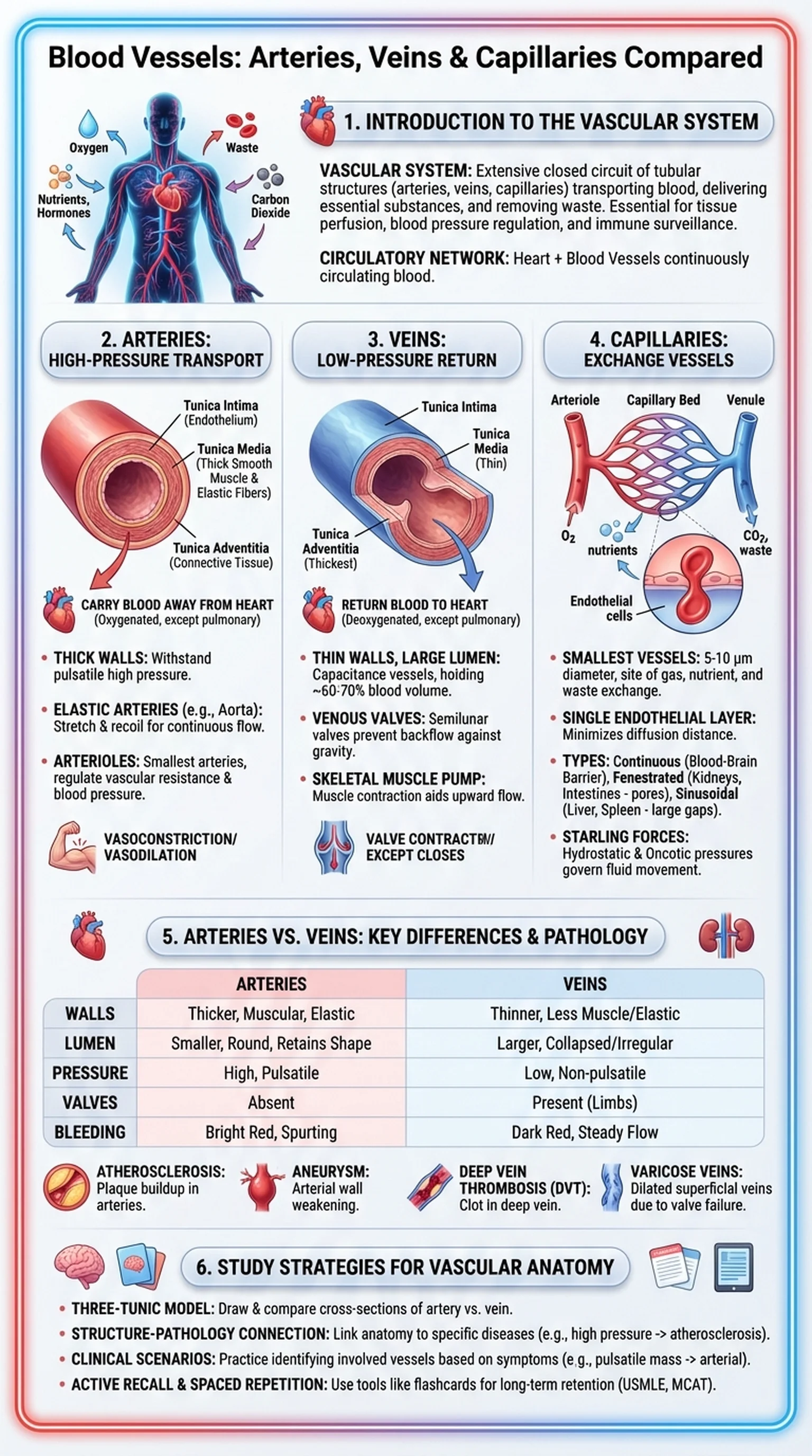
Blood vessels are the tubular structures that form the circulatory network responsible for transporting blood throughout the human body. Together, these vessels constitute the vascular system, an extensive closed circuit that delivers oxygen, nutrients, and hormones to tissues while removing metabolic waste products like carbon dioxide and urea. The vascular system is essential for maintaining tissue perfusion, regulating blood pressure, and enabling immune surveillance. Without a functioning network of blood vessels, no organ in the body could sustain its metabolic needs.

The vascular system is organized into three major categories of blood vessels: arteries, veins, and capillaries. Arteries carry oxygenated blood away from the heart to the tissues (with the exception of the pulmonary arteries, which carry deoxygenated blood to the lungs). Veins return deoxygenated blood from the tissues back to the heart (with the exception of the pulmonary veins). Capillaries are the microscopic vessels that connect the arterial and venous systems, forming the site where gas exchange, nutrient delivery, and waste removal actually occur.

Understanding blood vessels is foundational for students of anatomy, physiology, and clinical medicine. The structural differences between arteries, veins, and capillaries directly relate to their functional roles. Clinical conditions affecting blood vessels, including atherosclerosis, deep vein thrombosis, aneurysms, and varicose veins, are among the leading causes of morbidity and mortality worldwide. A strong grasp of vascular anatomy is therefore essential for diagnosing and managing cardiovascular disease.

Key Terms

Blood Vessels

Tubular structures including arteries, veins, and capillaries that form the circulatory network for transporting blood throughout the body.

Vascular System

The complete network of blood vessels in the body, including arteries, veins, and capillaries, responsible for circulating blood to all tissues.

Circulatory Network

The closed system of blood vessels and the heart that continuously circulates blood, delivering oxygen and nutrients while removing waste.

Perfusion

The passage of blood through the vascular system to an organ or tissue, ensuring adequate delivery of oxygen and nutrients.

Structure and Function of Arteries

Arteries are the high-pressure blood vessels that carry blood away from the heart and distribute it to the organs and tissues of the body. The walls of arteries are the thickest of all blood vessels, reflecting their need to withstand the pulsatile pressure generated by each heartbeat. Arterial walls are composed of three concentric layers, or tunics, each contributing to the vessel's structural integrity and functional properties.

The innermost layer, the tunica intima, consists of a single layer of endothelial cells resting on a thin basement membrane and a subendothelial layer of connective tissue. The endothelium plays a critical role in regulating vascular tone, preventing blood clotting, and mediating the exchange of substances between blood and tissue. The middle layer, the tunica media, is the thickest layer in arteries and is composed primarily of smooth muscle cells interspersed with elastic fibers. This layer enables arteries to constrict (vasoconstriction) and dilate (vasodilation), dynamically regulating blood flow and blood pressure. The outermost layer, the tunica adventitia (or tunica externa), is a connective tissue sheath containing collagen fibers, vasa vasorum (small blood vessels that supply the vessel wall itself), and nerve fibers.

Arteries are further classified by size and composition. Elastic arteries, such as the aorta and its major branches, have a high proportion of elastic fibers in the tunica media, allowing them to stretch during systole and recoil during diastole to maintain continuous blood flow. Muscular arteries, such as the radial and femoral arteries, have more smooth muscle and less elastin, enabling precise regulation of blood distribution to specific organs. Arterioles are the smallest arteries and serve as the primary site of vascular resistance, playing a crucial role in regulating blood pressure within the vascular system. When comparing arteries vs veins, the thicker walls and higher pressure environment of arteries are the most immediately distinguishing features.

Key Terms

Arteries

Thick-walled blood vessels that carry blood away from the heart under high pressure, distributing oxygenated blood to the body's tissues.

Tunica Media

The middle layer of the arterial wall, composed of smooth muscle and elastic fibers, responsible for vasoconstriction and vasodilation.

Elastic Arteries

Large arteries with a high proportion of elastic fibers that stretch and recoil with each heartbeat, such as the aorta.

Arterioles

The smallest arteries that serve as the main regulators of vascular resistance and blood pressure through smooth muscle contraction.

Structure and Function of Veins

Veins are the blood vessels that return deoxygenated blood from the peripheral tissues back to the heart. Compared to arteries, veins operate under significantly lower pressure and have evolved structural features that facilitate the return of blood against gravity, particularly from the lower extremities. The fundamental distinction in arteries vs veins lies in their wall structure: veins have thinner walls, larger lumens, and reduced smooth muscle and elastic tissue compared to their arterial counterparts.

Like arteries, veins possess three tunics, but the proportions differ markedly. The tunica intima of veins is similar to that of arteries, consisting of an endothelial lining on a basement membrane. However, the tunica media of veins is considerably thinner, containing less smooth muscle and fewer elastic fibers. The tunica adventitia is often the thickest layer in veins, composed of collagen and elastic fibers that provide structural support without the rigidity of arterial walls. This relatively thin-walled construction allows veins to act as capacitance vessels, capable of holding a large volume of blood. At any given time, approximately 60 to 70 percent of the body's blood volume resides in the venous system.

A defining feature of veins, especially in the limbs, is the presence of one-way venous valves. These semilunar valves prevent the backflow of blood, ensuring that venous return proceeds toward the heart despite the low-pressure environment. Venous return is further aided by the skeletal muscle pump, in which contraction of surrounding muscles compresses the veins and propels blood upward, and the respiratory pump, in which pressure changes during breathing create a pressure gradient that draws blood toward the thorax. Veins are classified by size: venules are the smallest veins that collect blood from capillary beds, medium veins contain well-developed valves, and large veins such as the superior and inferior vena cava return blood directly to the right atrium. Understanding how veins function within the vascular system is essential for recognizing conditions such as varicose veins, deep vein thrombosis, and chronic venous insufficiency.

Key Terms

Veins

Thin-walled blood vessels that return deoxygenated blood from peripheral tissues to the heart under low pressure.

Venous Valves

One-way semilunar valves within veins that prevent backflow of blood, ensuring unidirectional flow toward the heart.

Capacitance Vessels

A term for veins, reflecting their ability to hold a large proportion of the body's blood volume due to their distensible walls.

Skeletal Muscle Pump

The mechanism by which contraction of skeletal muscles surrounding veins compresses the vessels and assists venous return to the heart.

Capillaries: The Exchange Vessels

Capillaries are the smallest and most numerous blood vessels in the human body, forming vast networks called capillary beds that permeate virtually every tissue. With diameters of just 5 to 10 micrometers, barely wide enough for a single red blood cell to pass through, capillaries are the functional heart of the vascular system. It is here, and only here, that the actual exchange of gases, nutrients, and waste products occurs between the blood and the surrounding tissue cells. Every other type of blood vessel, whether artery or vein, exists ultimately to serve the capillary beds.

The structure of capillaries is elegantly simple. Their walls consist of a single layer of endothelial cells resting on a basement membrane, without the smooth muscle or elastic tissue found in arteries and veins. This thin wall minimizes the diffusion distance for gases and small molecules, enabling rapid exchange. The extremely slow flow velocity through capillaries (approximately 0.03 cm/sec) further facilitates exchange by maximizing the contact time between blood and tissue.

There are three main types of capillaries, each adapted to the specific needs of the tissue they serve. Continuous capillaries, found in the skin, lungs, and central nervous system, have tightly joined endothelial cells that restrict the passage of most molecules, forming the basis of the blood-brain barrier. Fenestrated capillaries, found in the kidneys, intestines, and endocrine glands, possess small pores (fenestrations) that allow faster exchange of fluids and small solutes. Sinusoidal capillaries (or discontinuous capillaries), found in the liver, spleen, and bone marrow, have large gaps between endothelial cells and an incomplete basement membrane, permitting the passage of large proteins and even whole cells. The movement of fluid across capillary walls is governed by Starling forces: hydrostatic pressure pushes fluid out of capillaries at the arterial end, while oncotic (osmotic) pressure draws fluid back in at the venous end. This balance ensures proper tissue hydration and is a critical concept in understanding how blood vessels maintain fluid homeostasis.

Key Terms

Capillaries

The smallest blood vessels, consisting of a single endothelial cell layer, where gas exchange, nutrient delivery, and waste removal occur between blood and tissues.

Fenestrated Capillaries

Capillaries with small pores in their endothelial walls that allow rapid exchange of fluids and solutes, found in kidneys, intestines, and endocrine organs.

Sinusoidal Capillaries

Large, irregularly shaped capillaries with gaps between endothelial cells, found in the liver, spleen, and bone marrow, allowing passage of large molecules and cells.

Starling Forces

The balance of hydrostatic and oncotic pressures across capillary walls that determines fluid movement between blood and interstitial tissue.

Arteries vs Veins: A Comprehensive Comparison

The comparison of arteries vs veins is one of the most fundamental topics in vascular anatomy and is frequently tested on board examinations. While both arteries and veins are blood vessels composed of three tunics, their structural and functional differences reflect their distinct roles within the vascular system. Understanding these differences is essential for clinical practice, as many diagnostic and therapeutic procedures depend on correctly identifying and accessing specific vessel types.

Structurally, arteries have thicker walls relative to their lumen size, with a prominently muscular and elastic tunica media. This thickness enables arteries to withstand the high pressure generated by ventricular contraction and to maintain their cylindrical shape even when empty, which is why arteries appear round in cross-section on histological slides. Veins, by contrast, have thinner walls and larger lumens, allowing them to accommodate a greater volume of blood at lower pressure. Veins typically appear collapsed or irregularly shaped in cross-section because their thin walls lack the rigidity to maintain a fixed shape without internal pressure.

Functionally, arteries carry blood away from the heart and distribute it at high pressure, while veins collect blood from capillary beds and return it to the heart at low pressure. The presence of venous valves in veins, absent in arteries, is a critical adaptation for low-pressure return. Arteries pulsate with each heartbeat, which is why clinicians can feel a pulse at superficial arterial sites (radial, carotid, femoral), whereas veins do not pulsate under normal conditions. When comparing arteries vs veins in the context of injury, arterial bleeding is bright red, spurting, and high-pressure, while venous bleeding is darker and flows steadily. Both arteries and veins are subject to disease: arteries develop atherosclerosis and aneurysms, while veins are prone to varicose formation and thrombosis. This comprehensive understanding of how blood vessels differ informs every aspect of cardiovascular diagnosis and treatment.

Key Terms

Arteries vs Veins

A fundamental anatomical comparison: arteries carry blood away from the heart under high pressure with thick walls, while veins return blood at low pressure with thinner walls and valves.

Atherosclerosis

A disease of arteries characterized by the buildup of lipid-rich plaques within the tunica intima, leading to vessel narrowing and increased risk of heart attack and stroke.

Deep Vein Thrombosis

The formation of a blood clot within a deep vein, most commonly in the lower extremities, which can embolize to the lungs causing pulmonary embolism.

Varicose Veins

Dilated, tortuous superficial veins caused by venous valve incompetence, most commonly occurring in the lower limbs.

Study Strategies for Blood Vessel Anatomy

Blood vessels and the vascular system are high-yield topics for medical board exams including the USMLE Step 1, COMLEX, and MCAT biology sections. The interplay between structural anatomy and clinical pathology makes this an area where systematic study pays significant dividends. Here are proven strategies for mastering this material.

First, learn the three-tunic model thoroughly and use it as a framework for comparison. Draw a cross-section of an artery and a vein side by side, labeling the tunica intima, tunica media, and tunica adventitia. Annotate the relative thickness of each layer and note the presence of valves in veins. This visual comparison of arteries vs veins helps cement the structural differences and makes it easy to predict functional differences on exam questions. Extend this comparison to include capillaries, noting that their walls consist of only an endothelial layer and basement membrane.

Second, connect structure to clinical pathology. For each vessel type, learn the most important associated diseases: atherosclerosis and aneurysms for arteries, deep vein thrombosis and varicose veins for veins, and edema for capillary dysfunction. Ask yourself how the structural features of each vessel type predispose it to its characteristic pathologies. For example, why does atherosclerosis affect arteries but not veins? The answer lies in the high-pressure environment and the interaction between blood flow dynamics and the arterial endothelium.

Third, use clinical scenarios to reinforce your knowledge of blood vessels. Practice identifying which vessels are affected in common presentations: a patient with a pulsatile abdominal mass likely has an aortic aneurysm (arterial), while a patient with leg swelling and pain after prolonged immobility likely has deep vein thrombosis (venous). Finally, use AI-powered study platforms like LectureScribe to generate flashcards, quizzes, and slide decks covering the vascular system, capillaries, and the comparison of arteries vs veins. Spaced repetition and active recall will help you retain the structural details and clinical correlations needed for exam success.

Key Terms

Active Recall

A study method involving actively retrieving information from memory, shown to improve long-term retention compared to passive review.

Spaced Repetition

A learning technique that schedules review sessions at progressively longer intervals to strengthen memory consolidation.

USMLE Step 1

A standardized medical licensing examination that tests foundational science concepts, including vascular anatomy and pathology.

Frequently Asked Questions

What are blood vessels and what do they do?

Blood vessels are tubular structures that transport blood throughout the body. They include arteries (carrying blood away from the heart), veins (returning blood to the heart), and capillaries (enabling exchange of gases, nutrients, and waste at the tissue level).

What is the difference between arteries and veins?

Arteries carry blood away from the heart under high pressure and have thick, muscular walls. Veins return blood to the heart under low pressure, have thinner walls, and contain one-way valves to prevent backflow. This arteries vs veins distinction is fundamental to vascular anatomy.

What are capillaries and why are they important?

Capillaries are the smallest blood vessels, with walls just one cell thick, where the actual exchange of oxygen, carbon dioxide, nutrients, and waste products occurs between blood and body tissues. They connect the arterial and venous systems.

What is the vascular system?

The vascular system is the complete network of blood vessels in the body, including arteries, veins, and capillaries. It works with the heart to circulate blood, delivering oxygen and nutrients to tissues while removing metabolic waste.

Why do veins have valves but arteries do not?

Veins operate under low pressure and must return blood to the heart against gravity, especially in the lower limbs. Venous valves prevent backflow and ensure unidirectional flow. Arteries do not need valves because the high pressure from the heart's pumping action maintains forward flow.

What are the three types of capillaries?

The three types are continuous capillaries (tightly sealed, found in the brain and skin), fenestrated capillaries (with small pores, found in kidneys and intestines), and sinusoidal capillaries (with large gaps, found in the liver and spleen).

What diseases affect blood vessels?

Major vascular diseases include atherosclerosis and aneurysms (affecting arteries), deep vein thrombosis and varicose veins (affecting veins), and edema resulting from capillary dysfunction. These conditions are among the leading causes of cardiovascular morbidity and mortality.

Study Anatomy Smarter

Upload your lecture notes and get AI-generated flashcards, quizzes, infographics, and study guides tailored to your curriculum.

Try LectureScribe Free

Create Study Materials

Transform your Anatomy lectures into flashcards, quizzes, and visual study guides with AI.

  • AI-generated flashcards & quizzes
  • Visual infographics from notes
  • Interactive study shorts
Start Studying Free

Quick Facts

SubjectAnatomy
Word Count2,500+
Slides5
InfographicIncluded
AuthorLectureScribe

Master Anatomy with LectureScribe

Upload your lecture notes and get AI-powered flashcards, quizzes, infographics, and study guides in minutes.