LectureScribe Logo
lecturescribe.io

LOCK IN VALENTINE PRICING BEFORE SALE ENDS!

Use code — New joiners get 40% OFF on annual plans

Plans will have higher prices after this sale ends

0days
:
0hours
:
0minutes
:
0seconds
Anatomy2,500+ words5 slides
🛡️

Lymphatic System: Function, Organs & Immune Role

Learn the lymphatic system including lymphatic function, lymph nodes, lymphatic organs, and lymphatic drainage. Understand how this system supports immunity and fluid balance.

L
LectureScribeAI-Powered Study Platform

Study Infographic

Complete lymphatic system overview illustrating lymph nodes, lymphatic organs, drainage pathways, and the integration of lymphatic function with immune defense.

Download
Lymphatic system diagram showing lymph nodes, lymphatic vessels, major lymphatic organs, and drainage patterns throughout the body

Interactive Study Short

Swipe through 5 slides about Lymphatic System: Function, Organs & Immune Role

1 / 5

Introduction to the Lymphatic System

The lymphatic system is a network of vessels, tissues, and organs that works in close partnership with the cardiovascular and immune systems to maintain fluid homeostasis, absorb dietary fats, and defend the body against infection. Despite being less familiar to many students than the circulatory or respiratory systems, the lymphatic system plays an indispensable role in human physiology. Without a functioning lymphatic system, tissues would become waterlogged with interstitial fluid, dietary lipids could not be properly absorbed, and the body's immune surveillance would be critically impaired.

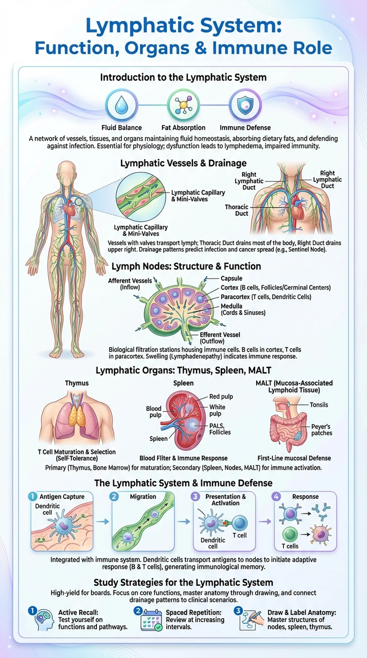
The lymphatic system can be understood as having three primary functions. First, lymphatic function in fluid balance involves the collection and return of excess interstitial fluid that leaks out of blood capillaries during normal circulation. This recovered fluid, once inside the lymphatic vessels, is called lymph. Approximately three liters of fluid are returned to the blood via the lymphatic system each day, preventing the accumulation of edema. Second, the lymphatic system serves as the primary route for absorbing long-chain fatty acids and fat-soluble vitamins from the small intestine through specialized lymphatic vessels called lacteals. Third, and perhaps most importantly, the lymphatic system is a central component of the adaptive and innate immune response, housing immune cells and providing the anatomical framework for immune surveillance and activation.

Understanding lymphatic function is essential for medical and anatomy students because dysfunction of this system underlies conditions ranging from lymphedema and infection to cancer metastasis. Lymphatic drainage patterns dictate how infections and malignancies spread through the body, making this knowledge directly applicable to clinical diagnosis and surgical planning.

Key Terms

Lymphatic System

A network of vessels, tissues, and organs that maintains fluid balance, absorbs dietary fats, and provides immune defense throughout the body.

Lymph

The clear, protein-rich fluid collected from interstitial spaces by lymphatic vessels and returned to the blood circulation.

Lymphatic Function

The combined roles of the lymphatic system in fluid recovery, fat absorption, and immune surveillance and response.

Lacteals

Specialized lymphatic capillaries in the villi of the small intestine that absorb dietary fats and transport them as chyle.

Lymphatic Vessels and Lymphatic Drainage

The lymphatic system begins with an extensive network of lymphatic capillaries, which are thin-walled, blind-ended vessels that permeate nearly every tissue in the body (with notable exceptions including the central nervous system, bone marrow, and avascular tissues like cartilage). Lymphatic capillaries are structurally designed to absorb interstitial fluid with remarkable efficiency. Their endothelial cells overlap loosely, forming one-way mini-valves that open when interstitial fluid pressure increases and close when internal pressure exceeds external pressure, preventing backflow.

Lymphatic capillaries merge to form larger collecting lymphatic vessels, which closely resemble veins in structure, possessing a similar three-tunic wall with smooth muscle and one-way valves that ensure unidirectional lymph flow. These collecting vessels pass through a series of lymph nodes, where lymph is filtered and immune cells survey its contents for pathogens and abnormal cells. The lymphatic drainage pattern of the body follows a predictable route: lymph from the collecting vessels converges into progressively larger trunks and ultimately empties into one of two main ducts.

The thoracic duct (left lymphatic duct) is the largest lymphatic vessel in the body and drains lymph from the left side of the head, left upper limb, left thorax, and the entire body below the diaphragm. It empties into the venous system at the junction of the left internal jugular and left subclavian veins. The right lymphatic duct drains the right side of the head, right upper limb, and right thorax, emptying at the corresponding right venous angle. Understanding lymphatic drainage is critical for predicting the spread of infections and metastatic cancer, as malignant cells often travel through lymphatic vessels to reach regional lymph nodes before disseminating further. Clinicians use knowledge of lymphatic drainage patterns to stage cancers, plan surgical lymph node dissections, and interpret sentinel lymph node biopsies.

Key Terms

Lymphatic Drainage

The pattern by which lymph flows through lymphatic vessels, nodes, trunks, and ducts before returning to the venous circulation.

Thoracic Duct

The largest lymphatic vessel, draining lymph from the left side of the body above the diaphragm and the entire body below the diaphragm into the left venous angle.

Collecting Lymphatic Vessels

Larger lymphatic vessels with valves and smooth muscle walls that transport lymph from capillaries through lymph nodes toward the lymphatic ducts.

Sentinel Lymph Node

The first lymph node to which cancer cells are most likely to spread from a primary tumor, used in staging and surgical decision-making.

Lymph Nodes: Structure and Function

Lymph nodes are small, bean-shaped lymphatic organs distributed along the course of lymphatic vessels throughout the body. There are approximately 600 to 700 lymph nodes in the human body, concentrated in regions such as the neck (cervical nodes), the armpit (axillary nodes), the groin (inguinal nodes), and the mesentery of the abdomen. Lymph nodes serve as biological filtration stations where lymph is screened for pathogens, foreign particles, and abnormal cells, making them a critical interface between the lymphatic system and the adaptive immune response.

The internal structure of lymph nodes is highly organized to maximize immune surveillance. Each node is surrounded by a fibrous capsule from which trabeculae extend inward, providing structural support. The cortex, located just beneath the capsule, contains densely packed lymphoid follicles composed primarily of B lymphocytes. Primary follicles contain naive B cells, while secondary follicles contain germinal centers where B cells are actively proliferating and differentiating into antibody-producing plasma cells in response to antigen stimulation. The paracortex, located between the cortex and the medulla, is rich in T lymphocytes and dendritic cells that present antigens to T cells, initiating cell-mediated immune responses. The medulla contains medullary cords of lymphoid tissue and medullary sinuses through which filtered lymph flows before exiting.

Lymph enters the node through multiple afferent lymphatic vessels on the convex surface, percolates through the cortical and medullary sinuses where it is filtered by resident macrophages and surveyed by lymphocytes, and exits through a single efferent lymphatic vessel at the hilum. When lymph nodes detect antigens, they mount an immune response that causes the node to enlarge, a condition clinically recognized as lymphadenopathy. Swollen lymph nodes are one of the most common physical examination findings and can indicate infection, autoimmune disease, or malignancy. The pattern of lymph node enlargement, combined with knowledge of lymphatic drainage territories, helps clinicians localize the source of pathology.

Key Terms

Lymph Nodes

Small, encapsulated lymphatic organs that filter lymph and house immune cells, serving as key sites for antigen detection and immune response initiation.

Germinal Center

A specialized area within lymph node follicles where B lymphocytes proliferate, undergo somatic hypermutation, and differentiate into plasma cells during an immune response.

Lymphadenopathy

Enlargement of lymph nodes, typically indicating an active immune response to infection, inflammation, or malignancy.

Afferent and Efferent Lymphatic Vessels

Afferent vessels bring lymph into a lymph node, while the single efferent vessel carries filtered lymph out of the node at the hilum.

Lymphatic Organs: Thymus, Spleen, and MALT

Beyond lymph nodes, the lymphatic system includes several major lymphatic organs that play distinct roles in immune cell development, maturation, and activation. These organs are classified as primary (central) or secondary (peripheral) lymphatic organs based on their function in the lifecycle of immune cells. The primary lymphatic organs are the bone marrow and the thymus, where lymphocytes are produced and mature. The secondary lymphatic organs include the spleen, lymph nodes, and mucosa-associated lymphoid tissue (MALT), where mature lymphocytes encounter antigens and mount immune responses.

The thymus is a bilobed organ located in the anterior mediastinum, superior to the heart. It is the site where T lymphocytes (T cells) mature and undergo selection. During thymic education, developing T cells that react too strongly to self-antigens are eliminated (negative selection), while those that can recognize self-MHC molecules are preserved (positive selection). This process ensures self-tolerance and prevents autoimmune disease. The thymus is largest and most active during childhood and progressively involutes (shrinks) after puberty, being largely replaced by adipose tissue in older adults.

The spleen is the largest lymphatic organ, located in the left upper quadrant of the abdomen. It functions as a blood filter rather than a lymph filter, removing old or damaged red blood cells (red pulp function) and mounting immune responses against blood-borne antigens (white pulp function). The white pulp contains periarteriolar lymphoid sheaths (PALS) rich in T cells and follicles containing B cells, mirroring the organization of lymph nodes. MALT encompasses all lymphoid tissue associated with mucosal surfaces, including the tonsils, Peyer's patches in the small intestine, and the appendix. These lymphatic organs provide first-line immune defense at the body's mucosal barriers, where most pathogens enter. Together, the lymphatic organs form an integrated network that ensures comprehensive immune surveillance throughout the body.

Key Terms

Lymphatic Organs

Organs of the lymphatic system including the thymus, spleen, lymph nodes, and MALT, each serving specialized roles in immune cell development or activation.

Thymus

A primary lymphatic organ in the mediastinum where T lymphocytes mature and undergo positive and negative selection to ensure self-tolerance.

Spleen

The largest secondary lymphatic organ, responsible for filtering blood, recycling old red blood cells, and mounting immune responses against blood-borne pathogens.

MALT

Mucosa-associated lymphoid tissue, including tonsils and Peyer's patches, that provides immune defense at mucosal surfaces where pathogens commonly enter the body.

The Lymphatic System and Immune Defense

The lymphatic system is inseparable from the immune system, providing the structural framework through which immune cells travel, encounter antigens, and coordinate defensive responses. Lymphatic function in immunity involves both the innate immune response, which provides rapid but nonspecific defense, and the adaptive immune response, which generates highly specific, long-lasting protection against pathogens.

Within the lymphatic system, dendritic cells in peripheral tissues capture antigens from invading pathogens and travel through afferent lymphatic vessels to regional lymph nodes. There, they present processed antigen fragments to naive T cells in the paracortex, initiating the adaptive immune response. Helper T cells (CD4+) coordinate the response by activating B cells in the follicles and stimulating cytotoxic T cells (CD8+) that can directly kill infected cells. Activated B cells in the germinal centers of lymph nodes undergo clonal expansion and affinity maturation, producing high-affinity antibodies that are released into the lymph and blood to neutralize pathogens. Memory B and T cells generated during this response persist in lymphatic organs, providing the basis for immunological memory and faster responses upon re-exposure.

The lymphatic system also plays a critical role in immune tolerance and the prevention of autoimmune disease. The thymus eliminates self-reactive T cells during development, while regulatory T cells circulating through the lymphatic system suppress inappropriate immune responses in the periphery. Dysfunction of the lymphatic system can lead to immunodeficiency, chronic infection, or autoimmune disorders. Furthermore, the lymphatic system is a pathway for cancer metastasis: tumor cells can enter lymphatic vessels, travel to regional lymph nodes, and establish secondary tumors. This is why lymph node status is a critical factor in cancer staging and prognosis, and why understanding lymphatic drainage patterns is essential for surgical oncology.

Key Terms

Adaptive Immune Response

The branch of immunity involving antigen-specific T and B lymphocytes that provides targeted defense and immunological memory.

Dendritic Cells

Antigen-presenting cells that capture pathogens in peripheral tissues and migrate through lymphatic vessels to lymph nodes to activate T cells.

Immunological Memory

The ability of the immune system to respond more rapidly and effectively to previously encountered antigens, mediated by memory B and T cells in lymphatic organs.

Cancer Metastasis via Lymphatics

The spread of malignant cells through lymphatic vessels to regional lymph nodes and beyond, a key factor in cancer staging and treatment planning.

Study Strategies for the Lymphatic System

The lymphatic system is a high-yield topic for medical board exams including the USMLE Step 1, COMLEX, and MCAT biology sections. Its integration with immunology makes it one of the most frequently tested areas in both anatomy and pathology. Here are effective strategies for mastering this material.

First, organize your study around the three core functions of the lymphatic system: fluid balance, fat absorption, and immune defense. For each function, trace the anatomical pathway involved. For fluid balance, follow the journey of interstitial fluid as it enters lymphatic capillaries, travels through collecting vessels and lymph nodes, and returns to the blood via the thoracic duct. For fat absorption, understand how lacteals in the intestinal villi absorb chylomicrons and transport them through the lymphatic system. For immune defense, learn how antigens travel from peripheral tissues through lymphatic vessels to lymph nodes, where they activate B and T cells.

Second, master the anatomy of lymph nodes and lymphatic organs by drawing and labeling them from memory. For a lymph node, sketch the capsule, cortex with follicles, paracortex, medulla, afferent and efferent vessels, and hilum. Label where B cells, T cells, and macrophages are concentrated. For the spleen, distinguish between red pulp and white pulp and their respective functions. For the thymus, understand positive and negative selection. This structural knowledge forms the foundation for understanding how the lymphatic system mounts immune responses.

Third, learn lymphatic drainage patterns and connect them to clinical scenarios. Know which body regions drain to which lymph node groups (axillary, inguinal, cervical, mediastinal) and understand how lymphatic drainage determines the spread of cancer. For example, breast cancer commonly metastasizes first to axillary lymph nodes because the breast drains primarily to this group. Finally, use AI-powered platforms like LectureScribe to generate flashcards, practice questions, and slide decks covering lymph nodes, lymphatic organs, lymphatic function, and lymphatic drainage. Spaced repetition and active recall will help you retain the integrated anatomical and immunological knowledge needed for exam success.

Key Terms

Active Recall

A learning technique that involves actively retrieving information from memory rather than passively reviewing notes, proven to strengthen long-term retention.

Spaced Repetition

A study strategy that schedules review at progressively increasing intervals to optimize memory consolidation over time.

USMLE Step 1

A standardized medical licensing examination that tests foundational science knowledge, including extensive lymphatic system and immunology content.

Frequently Asked Questions

What is the lymphatic system and what does it do?

The lymphatic system is a network of vessels, nodes, and organs that maintains fluid balance by recovering excess interstitial fluid, absorbs dietary fats from the intestines, and provides immune defense by housing and activating immune cells throughout the body.

What are lymph nodes and why do they swell?

Lymph nodes are small, bean-shaped organs that filter lymph and house immune cells. They swell (lymphadenopathy) when immune cells within them proliferate in response to infection, inflammation, or the presence of cancer cells, indicating an active immune response.

What is lymphatic function in the immune system?

Lymphatic function in immunity includes transporting antigens and antigen-presenting cells to lymph nodes, providing the environment for B and T cell activation, generating antibodies and memory cells, and circulating immune cells throughout the body for surveillance.

What are the major lymphatic organs?

The major lymphatic organs include the thymus (T cell maturation), spleen (blood filtration and immune response), lymph nodes (lymph filtration and immune activation), and MALT including tonsils and Peyer's patches (mucosal immune defense).

What is lymphatic drainage and why is it clinically important?

Lymphatic drainage refers to the pathways through which lymph flows from tissues through lymphatic vessels and nodes back to the bloodstream. It is clinically important because cancer cells and infections spread along these drainage routes, making knowledge of drainage patterns essential for cancer staging and treatment.

What happens when the lymphatic system is damaged?

Damage to the lymphatic system can cause lymphedema (tissue swelling from fluid accumulation), increased susceptibility to infections due to impaired immune surveillance, and disruption of fat absorption. Surgical removal of lymph nodes during cancer treatment is a common cause of lymphedema.

How does the lymphatic system relate to cancer spread?

Malignant tumor cells can enter lymphatic vessels and travel to regional lymph nodes, where they may establish secondary tumors (metastases). The status of lymph nodes is a key factor in cancer staging, and sentinel lymph node biopsy is used to determine whether cancer has begun to spread.

Study Anatomy Smarter

Upload your lecture notes and get AI-generated flashcards, quizzes, infographics, and study guides tailored to your curriculum.

Try LectureScribe Free

Create Study Materials

Transform your Anatomy lectures into flashcards, quizzes, and visual study guides with AI.

  • AI-generated flashcards & quizzes
  • Visual infographics from notes
  • Interactive study shorts
Start Studying Free

Quick Facts

SubjectAnatomy
Word Count2,500+
Slides5
InfographicIncluded
AuthorLectureScribe

Master Anatomy with LectureScribe

Upload your lecture notes and get AI-powered flashcards, quizzes, infographics, and study guides in minutes.