LectureScribe Logo
lecturescribe.io

LOCK IN VALENTINE PRICING BEFORE SALE ENDS!

Use code — New joiners get 40% OFF on annual plans

Plans will have higher prices after this sale ends

0days
:
0hours
:
0minutes
:
0seconds
Biology2,500+ words5 slides
🔄

Cell Cycle and Mitosis Phases: Complete Study Guide

Master the cell cycle and mitosis phases with this complete study guide. Learn interphase, cell division stages, and cytokinesis with clear explanations and practice questions.

L
LectureScribeAI-Powered Study Platform

Study Infographic

Complete cell cycle and mitosis phases diagram illustrating interphase preparation, nuclear division stages, and cytokinesis.

Download
Cell cycle diagram showing interphase stages G1, S, and G2 followed by the mitosis phases prophase, metaphase, anaphase, telophase, and cytokinesis

Interactive Study Short

Swipe through 5 slides about Cell Cycle and Mitosis Phases: Complete Study Guide

1 / 5

What Is the Cell Cycle?

The cell cycle is the ordered sequence of events that a cell undergoes from its formation to the moment it divides into two daughter cells. This fundamental biological process ensures that organisms can grow, repair damaged tissues, and reproduce. Every living cell, from single-celled bacteria to the trillions of cells in the human body, follows some version of the cell cycle. Understanding the cell cycle is essential for fields ranging from developmental biology to cancer research, where uncontrolled cell division drives tumor formation.

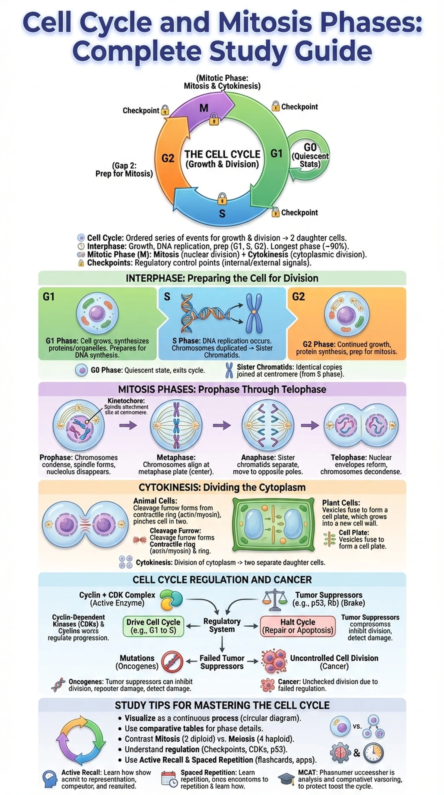
The cell cycle is broadly divided into two major phases: interphase and the mitotic phase (M phase). Interphase is the longest portion of the cell cycle, occupying roughly 90 percent of the total cycle time in a typical mammalian cell. During interphase, the cell grows in size, duplicates its organelles, and replicates its entire genome in preparation for division. Interphase itself is subdivided into three distinct stages: G1 (Gap 1), S (Synthesis), and G2 (Gap 2). Each of these stages serves a specific preparatory function that ensures the cell is ready to enter the mitotic phase.

The mitotic phase encompasses both mitosis, the division of the nucleus, and cytokinesis, the division of the cytoplasm. Together, these processes produce two genetically identical daughter cells. The cell cycle is tightly regulated by a system of checkpoints and regulatory proteins, including cyclins and cyclin-dependent kinases, that monitor cell size, DNA integrity, and external growth signals. When these regulatory mechanisms fail, the result can be uncontrolled cell division, a hallmark of cancer. Mastering the stages of the cell cycle provides a critical foundation for understanding how organisms develop and how diseases related to cell division arise.

Key Terms

Cell Cycle

The ordered series of events involving cell growth and cell division that produces two new daughter cells.

Interphase

The phase of the cell cycle during which the cell grows, replicates its DNA, and prepares for division; includes G1, S, and G2 stages.

Mitotic Phase

The phase of the cell cycle that includes mitosis (nuclear division) and cytokinesis (cytoplasmic division).

Checkpoints

Regulatory control points in the cell cycle where the cell evaluates internal and external signals before proceeding to the next phase.

Interphase: Preparing the Cell for Division

Interphase is often mischaracterized as a resting phase, but in reality it is the most metabolically active period of the cell cycle. During interphase, the cell is carrying out its normal functions while simultaneously preparing for cell division. The three sub-stages of interphase, G1, S, and G2, each play a crucial role in ensuring that the cell is fully equipped to divide successfully.

During the G1 phase (Gap 1), the cell grows in size, synthesizes proteins and organelles, and carries out its specialized functions. This is typically the longest phase of interphase and represents the primary period of cell growth. The cell also evaluates whether conditions are favorable for division. At the end of G1, the cell encounters the G1 checkpoint (also known as the restriction point in mammalian cells), where it assesses nutrient availability, cell size, and growth factor signals. If conditions are not met, the cell may exit the cell cycle and enter a quiescent state called G0.

The S phase (Synthesis) is when DNA replication occurs. Each chromosome is duplicated to produce two identical sister chromatids joined at the centromere. Histone proteins are also synthesized during S phase to package the newly replicated DNA. The centrosome, which will organize the mitotic spindle, is also duplicated during this stage. Following DNA replication, the cell enters the G2 phase (Gap 2), a shorter growth period during which the cell continues to produce proteins and organelles needed for mitosis. The G2 checkpoint verifies that DNA replication has been completed accurately and that any damage has been repaired. Once the cell passes the G2 checkpoint, it is committed to entering the mitotic phase and beginning cell division.

Key Terms

G1 Phase

The first gap phase of interphase during which the cell grows in size and prepares for DNA synthesis.

S Phase

The synthesis phase of interphase in which the cell replicates its entire genome, producing sister chromatids.

G2 Phase

The second gap phase of interphase during which the cell continues to grow and prepares for mitosis.

Sister Chromatids

Two identical copies of a single chromosome that are joined at the centromere, formed during DNA replication in S phase.

G0 Phase

A quiescent state in which the cell exits the cell cycle and ceases to divide, often permanently in highly specialized cells.

Mitosis Phases: Prophase Through Telophase

Mitosis is the process of nuclear division that distributes duplicated chromosomes equally between two daughter nuclei. The mitosis phases are traditionally divided into four sequential stages: prophase, metaphase, anaphase, and telophase. Some textbooks also recognize prometaphase as a distinct stage between prophase and metaphase. Understanding each of these mitosis phases in detail is essential for biology students at every level.

During prophase, the first stage of mitosis, the chromatin condenses into visible chromosomes. Each chromosome consists of two sister chromatids joined at the centromere. The mitotic spindle begins to form as centrosomes migrate toward opposite poles of the cell, and the nucleolus disappears. In prometaphase, the nuclear envelope breaks down, and spindle fibers attach to the kinetochores, specialized protein structures located at the centromere of each chromosome. This connection is critical for chromosome movement.

Metaphase is characterized by the alignment of all chromosomes along the metaphase plate, an imaginary equatorial plane midway between the two spindle poles. The spindle assembly checkpoint ensures that every kinetochore is properly attached to spindle fibers before the cell proceeds. This checkpoint is a vital safeguard against aneuploidy, the condition of having an abnormal number of chromosomes. During anaphase, the cohesin proteins holding sister chromatids together are cleaved by the enzyme separase, and the chromatids are pulled toward opposite poles of the cell by shortening spindle fibers.

In telophase, the final stage of mitosis, the separated chromosomes arrive at opposite poles and begin to decondense back into chromatin. A nuclear envelope reforms around each set of chromosomes, nucleoli reappear, and the spindle apparatus disassembles. At the completion of telophase, the cell contains two genetically identical nuclei, setting the stage for cytokinesis to physically divide the cell into two separate daughter cells.

Key Terms

Prophase

The first phase of mitosis during which chromosomes condense, the spindle begins to form, and the nucleolus disappears.

Metaphase

The phase of mitosis in which chromosomes align along the metaphase plate at the center of the cell.

Anaphase

The phase of mitosis during which sister chromatids separate and move toward opposite poles of the cell.

Telophase

The final phase of mitosis in which chromosomes decondense, nuclear envelopes reform, and the mitotic spindle disassembles.

Kinetochore

A protein complex at the centromere of a chromosome where spindle fibers attach during mitosis.

Cytokinesis: Dividing the Cytoplasm

Cytokinesis is the final step of cell division, during which the cytoplasm of the parent cell is divided to produce two separate daughter cells. While mitosis distributes the nuclear contents, cytokinesis ensures that each daughter cell receives an adequate share of organelles, cytoplasm, and cell membrane. In most cells, cytokinesis begins during late anaphase or telophase and overlaps with the final stages of mitosis, but it is technically a distinct process.

In animal cells, cytokinesis occurs through a mechanism known as cleavage furrow formation. A contractile ring composed of actin filaments and myosin motor proteins assembles just beneath the plasma membrane at the cell's equator. This ring contracts inward, pinching the cell progressively until the two daughter cells separate completely. The position of the cleavage furrow is determined by signals from the mitotic spindle, ensuring that the division plane corresponds to the metaphase plate.

In plant cells, cytokinesis proceeds differently because the rigid cell wall prevents cleavage furrow formation. Instead, vesicles derived from the Golgi apparatus accumulate along the plane of division and fuse to form a structure called the cell plate. The cell plate grows outward from the center of the cell toward the existing cell walls, eventually forming a new cell wall that separates the two daughter cells. Each daughter cell then synthesizes additional cell wall material and plasma membrane to complete the separation.

The successful completion of cytokinesis marks the end of cell division and the beginning of a new cell cycle for each daughter cell. Errors during cytokinesis can result in binucleated cells or cells with abnormal amounts of cytoplasm. In some specialized contexts, such as the formation of muscle fibers, cytokinesis is deliberately suppressed, producing large multinucleated cells called syncytia. Understanding how cytokinesis works alongside the mitosis phases provides a complete picture of how one cell becomes two.

Key Terms

Cytokinesis

The process of cytoplasmic division that follows mitosis, resulting in two separate daughter cells.

Cleavage Furrow

An indentation in the plasma membrane of an animal cell formed by a contractile ring during cytokinesis.

Cell Plate

A structure formed during cytokinesis in plant cells from fused Golgi vesicles, which develops into a new cell wall.

Contractile Ring

A ring of actin and myosin filaments that contracts during cytokinesis to pinch the animal cell in two.

Cell Cycle Regulation and Cancer

The cell cycle is governed by an intricate regulatory system that ensures cells divide only when appropriate. At the core of this system are cyclin-dependent kinases (CDKs), enzymes that are activated when they bind to regulatory proteins called cyclins. Different cyclin-CDK complexes control the transitions between specific phases of the cell cycle. For example, the cyclin D-CDK4/6 complex drives progression through the G1 phase, while the cyclin B-CDK1 complex triggers entry into mitosis.

Cell division is also regulated by tumor suppressor genes and proto-oncogenes. Tumor suppressors, such as p53 and Rb (retinoblastoma protein), act as brakes on the cell cycle. The p53 protein is often called the "guardian of the genome" because it halts the cell cycle when DNA damage is detected, allowing time for repair or triggering apoptosis (programmed cell death) if the damage is irreparable. Proto-oncogenes encode proteins that normally promote cell growth and division; however, when mutated into oncogenes, they can drive excessive cell division independent of normal regulatory signals.

When the regulatory machinery of the cell cycle breaks down, the result is often cancer. Cancer cells characteristically bypass checkpoints, ignore growth-inhibitory signals, and continue through the cell cycle unchecked. Mutations in p53 are found in over half of all human cancers, underscoring its critical role. Understanding cell cycle regulation has led to the development of targeted cancer therapies, including CDK inhibitors that specifically block the kinases driving tumor cell division.

For students preparing for exams, the connection between the cell cycle and cancer is a high-yield topic. Knowing how cyclins, CDKs, tumor suppressors, and oncogenes interact provides insight not only into normal cell division but also into the molecular basis of one of the most significant diseases in medicine.

Key Terms

Cyclin-Dependent Kinases (CDKs)

Enzymes that regulate cell cycle progression when activated by binding to cyclin proteins.

Tumor Suppressors

Genes or proteins, such as p53 and Rb, that inhibit cell division and prevent uncontrolled growth.

Oncogenes

Mutated forms of proto-oncogenes that promote uncontrolled cell division and can lead to cancer.

Apoptosis

Programmed cell death triggered by internal or external signals, serving as a safeguard against damaged or unnecessary cells.

Study Tips for Mastering the Cell Cycle and Mitosis

The cell cycle and mitosis phases are among the most tested topics in introductory biology, AP Biology, and the MCAT. Developing a deep understanding requires strategies that move beyond simple memorization of phase names and into a functional understanding of why each stage matters.

First, learn the cell cycle as a continuous process rather than a set of isolated stages. Create a circular diagram that flows from G1 through S, G2, and the mitosis phases (prophase, metaphase, anaphase, telophase), ending with cytokinesis. Annotate each stage with its key events and checkpoints. This visual approach helps you see how interphase prepares the cell for cell division and how each checkpoint guards the transition to the next phase.

Second, use comparative tables to distinguish the mitosis phases from one another. For each phase, note what happens to the chromosomes, the spindle, the nuclear envelope, and the cell membrane. This structured approach makes it easy to answer identification questions where you are given a cell image and asked to name the stage. Third, pay special attention to the differences between mitosis and meiosis. While this guide focuses on mitosis, exam questions frequently require students to compare the two processes. Mitosis produces two identical diploid daughter cells, while meiosis produces four genetically unique haploid cells.

Fourth, understand regulation. Know the three major checkpoints (G1, G2, and spindle assembly), the roles of cyclins and CDKs, and how tumor suppressors like p53 function. These concepts are essential for connecting the cell cycle to cancer biology. Finally, leverage technology for active study. Platforms like LectureScribe can transform your lecture notes on cell division into interactive slide decks, flashcards, and quiz questions, helping you practice active recall and spaced repetition for long-term mastery of the cell cycle and its phases.

Key Terms

Active Recall

A study technique in which learners actively retrieve information from memory rather than passively reviewing notes.

Spaced Repetition

A learning strategy that involves reviewing material at increasing intervals to strengthen long-term memory retention.

MCAT

The Medical College Admission Test; a standardized exam required for admission to medical schools in the United States and Canada.

Frequently Asked Questions

What are the phases of the cell cycle?

The cell cycle consists of interphase (G1, S, and G2 phases) and the mitotic phase (M phase), which includes mitosis and cytokinesis. During interphase, the cell grows and replicates its DNA. During the mitotic phase, the cell divides its nucleus and cytoplasm to produce two daughter cells.

What are the four mitosis phases in order?

The four mitosis phases in order are prophase, metaphase, anaphase, and telophase. Some textbooks also include prometaphase as a distinct stage between prophase and metaphase. Each phase involves specific changes to the chromosomes, spindle, and nuclear envelope.

What happens during interphase?

During interphase, the cell grows in size (G1), replicates its DNA (S phase), and prepares for mitosis (G2). Interphase is the longest part of the cell cycle, accounting for about 90 percent of total cycle time, and is essential for ensuring the cell is ready for division.

What is cytokinesis and when does it occur?

Cytokinesis is the division of the cytoplasm that follows mitosis, producing two separate daughter cells. In animal cells, it occurs via cleavage furrow formation, while in plant cells it involves the formation of a cell plate. Cytokinesis typically begins during late anaphase or telophase.

How does cell division relate to cancer?

Cancer results from uncontrolled cell division caused by mutations in genes that regulate the cell cycle. When tumor suppressors like p53 are inactivated or proto-oncogenes are mutated into oncogenes, cells bypass normal checkpoints and divide without restraint, forming tumors.

What is the difference between mitosis and cell division?

Mitosis is specifically the division of the cell nucleus into two identical nuclei. Cell division is the broader term that includes both mitosis (nuclear division) and cytokinesis (cytoplasmic division). Together, these two processes produce two complete daughter cells.

What are the checkpoints of the cell cycle?

The three major cell cycle checkpoints are the G1 checkpoint (restriction point), the G2 checkpoint, and the spindle assembly checkpoint during metaphase. These checkpoints ensure the cell has adequate size, intact DNA, and proper spindle attachment before proceeding to the next phase.

Study Biology Smarter

Upload your lecture notes and get AI-generated flashcards, quizzes, infographics, and study guides tailored to your curriculum.

Try LectureScribe Free

Create Study Materials

Transform your Biology lectures into flashcards, quizzes, and visual study guides with AI.

  • AI-generated flashcards & quizzes
  • Visual infographics from notes
  • Interactive study shorts
Start Studying Free

Quick Facts

SubjectBiology
Word Count2,500+
Slides5
InfographicIncluded
AuthorLectureScribe

Master Biology with LectureScribe

Upload your lecture notes and get AI-powered flashcards, quizzes, infographics, and study guides in minutes.