LectureScribe Logo
lecturescribe.io

LOCK IN VALENTINE PRICING BEFORE SALE ENDS!

Use code — New joiners get 40% OFF on annual plans

Plans will have higher prices after this sale ends

0days
:
0hours
:
0minutes
:
0seconds
Biology3,000+ words5 slides
🧬

Mitosis vs Meiosis: Key Differences, Comparison Table, and Stages Explained

Compare and contrast meiosis and mitosis with clear explanations, a comparison table, and the 3 differences between mitosis and meiosis every student must know.

L
LectureScribeAI-Powered Study Platform

Study Infographic

Side-by-side comparison between mitosis and meiosis illustrating stages, daughter cell outcomes, and genetic variation.

Download
Mitosis vs meiosis comparison diagram showing stages, chromosome numbers, and key differences between mitosis and meiosis

Interactive Study Short

Swipe through 5 slides about Mitosis vs Meiosis: Key Differences, Comparison Table, and Stages Explained

1 / 5

What Are Mitosis and Meiosis?

Mitosis and meiosis are the two fundamental types of cell division in eukaryotic organisms. While both processes involve the replication and segregation of genetic material, they serve fundamentally different biological purposes and produce very different outcomes. Understanding these two processes is essential for students of biology, genetics, and medicine, as the difference between mitosis and meiosis underpins everything from tissue growth to genetic diversity.

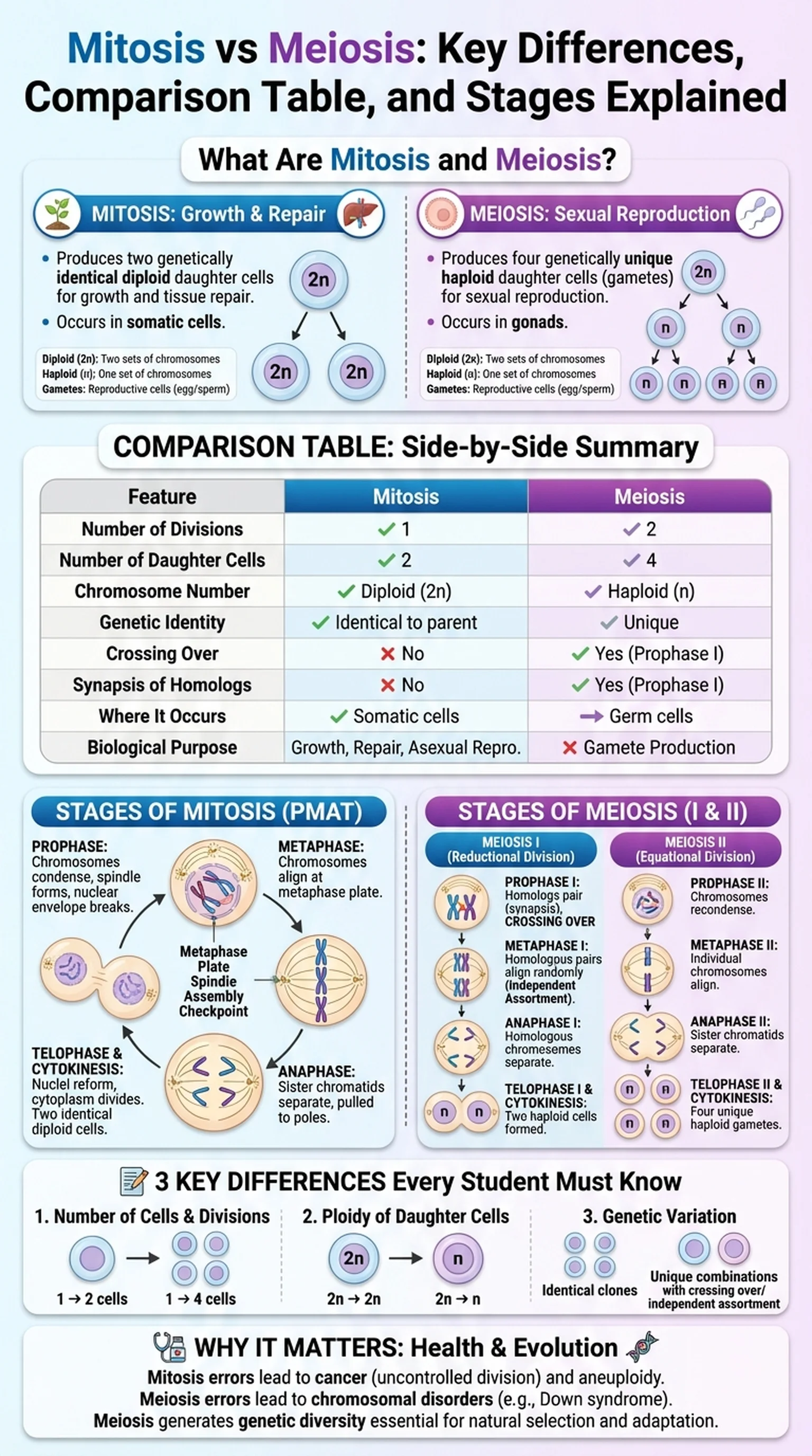
Mitosis is the process of cell division that produces two genetically identical daughter cells, each containing the same number of chromosomes as the original parent cell. It is the mechanism by which organisms grow, repair damaged tissues, and maintain their body structures. Every time a skin cell divides to replace a lost cell, or a liver cell replicates during organ regeneration, it does so through mitosis. The result is always two diploid cells that are genetic clones of the parent.

Meiosis, by contrast, is a specialized form of cell division that produces four genetically unique daughter cells, each containing half the number of chromosomes found in the parent cell. Meiosis occurs exclusively in the gonads (ovaries and testes in animals) and is the process that generates gametes: eggs and sperm. The reduction in chromosome number during meiosis is critical because it ensures that when two gametes fuse during fertilization, the resulting zygote has the correct diploid chromosome count. The meiosis and mitosis difference in purpose, therefore, is straightforward: mitosis is for growth and repair, while meiosis is for sexual reproduction.

Key Terms

Mitosis

A type of cell division that produces two genetically identical diploid daughter cells from a single parent cell.

Meiosis

A type of cell division that produces four genetically unique haploid daughter cells (gametes) from a single diploid parent cell.

Diploid

A cell containing two complete sets of chromosomes (2n), one inherited from each parent.

Haploid

A cell containing a single set of chromosomes (n), as found in gametes.

Gametes

Reproductive cells (eggs and sperm) produced by meiosis that carry half the genetic information of the organism.

Key Differences Between Mitosis and Meiosis

The difference between meiosis and mitosis extends across multiple dimensions, including the number of divisions, the genetic composition of daughter cells, the role of crossing over, and the biological context in which each process occurs. Students who can clearly articulate these differences demonstrate a strong foundation in cell biology.

First, consider the number of cell divisions. Mitosis involves a single round of division, producing two daughter cells. Meiosis involves two successive rounds of division, meiosis I and meiosis II, producing a total of four daughter cells. This fundamental structural difference between mitosis and meiosis accounts for the reduction in chromosome number during meiosis.

Second, the genetic outcome differs dramatically. In mitosis, the daughter cells are genetically identical to the parent cell and to each other. In meiosis, the daughter cells are genetically unique due to two mechanisms: crossing over (recombination) during prophase I and independent assortment of homologous chromosomes during metaphase I. These processes shuffle genetic material, ensuring that no two gametes are alike.

Third, the chromosome number in the daughter cells is different. Mitosis maintains the diploid (2n) chromosome count, so a human cell with 46 chromosomes produces two cells each with 46 chromosomes. Meiosis reduces the chromosome count to haploid (n), so a human cell with 46 chromosomes produces four cells each with 23 chromosomes. This is perhaps the most frequently tested meiosis and mitosis difference on biology exams.

Fourth, the biological roles differ. Mitosis drives growth, tissue repair, and asexual reproduction in some organisms. Meiosis is dedicated solely to producing gametes for sexual reproduction. Finally, homologous chromosome pairing (synapsis) occurs only in meiosis I, not in mitosis. During synapsis, homologous chromosomes align side by side, enabling crossing over. This event is entirely absent from mitosis, making it one of the clearest ways to compare meiosis and mitosis at the mechanistic level.

Key Terms

Crossing Over

The exchange of genetic material between homologous chromosomes during prophase I of meiosis, generating genetic diversity.

Independent Assortment

The random orientation of homologous chromosome pairs during metaphase I, leading to varied combinations of maternal and paternal chromosomes in gametes.

Synapsis

The pairing of homologous chromosomes during prophase I of meiosis, facilitated by the synaptonemal complex.

Homologous Chromosomes

A pair of chromosomes, one from each parent, that have the same genes at the same loci but may carry different alleles.

Recombination

The process by which DNA segments are exchanged between homologous chromosomes, creating new allele combinations.

Comparison Between Mitosis and Meiosis: Side-by-Side Table

One of the most effective ways to study cell division is to organize the comparison between mitosis and meiosis in a structured table format. A difference between mitosis and meiosis table allows you to see all the contrasts at a glance and is frequently used in textbooks and on exams.

Number of Divisions: Mitosis has one division; Meiosis has two divisions (meiosis I and meiosis II). Number of Daughter Cells: Mitosis produces two; Meiosis produces four. Chromosome Number in Daughter Cells: Mitosis maintains diploid (2n); Meiosis reduces to haploid (n). Genetic Identity: Mitosis produces identical cells; Meiosis produces genetically unique cells. Crossing Over: Does not occur in mitosis; Occurs during prophase I of meiosis. Synapsis of Homologs: Does not occur in mitosis; Occurs during prophase I of meiosis. Where It Occurs: Mitosis occurs in somatic (body) cells; Meiosis occurs in germ cells of the gonads. Biological Purpose: Mitosis is for growth and repair; Meiosis is for gamete production. Sister Chromatid Separation: Occurs in anaphase of mitosis; Occurs in anaphase II of meiosis. Homologous Chromosome Separation: Does not apply to mitosis; Occurs in anaphase I of meiosis.

When you compare and contrast meiosis and mitosis using this table format, several patterns emerge. Both processes share the same basic stages (prophase, metaphase, anaphase, telophase), but meiosis runs through these stages twice. Both require DNA replication during S phase before division begins. However, the comparison between mitosis and meiosis reveals that only meiosis introduces genetic variation through crossing over and independent assortment. This structured comparison is invaluable for exam preparation, particularly for standardized tests like the AP Biology exam and the MCAT, where the difference between mitosis and meiosis table format questions are common.

Key Terms

Somatic Cells

All body cells except gametes; they divide by mitosis and are diploid.

Germ Cells

Cells in the gonads that undergo meiosis to produce haploid gametes.

Sister Chromatids

Two identical copies of a chromosome joined at the centromere, formed during DNA replication.

S Phase

The phase of the cell cycle during which DNA is replicated in preparation for cell division.

Stages of Mitosis: Prophase, Metaphase, Anaphase, and Telophase

Mitosis proceeds through four main stages, often remembered by the mnemonic PMAT: Prophase, Metaphase, Anaphase, and Telophase. Each stage is characterized by specific chromosomal and cellular events that ensure the accurate distribution of genetic material to daughter cells.

During prophase, the chromatin condenses into visible chromosomes, each consisting of two sister chromatids joined at the centromere. The mitotic spindle begins to form from the centrosomes, which migrate to opposite poles of the cell. The nuclear envelope breaks down, allowing spindle fibers to access the chromosomes. In some descriptions, prometaphase is identified as a substage where kinetochore microtubules attach to the kinetochores on each chromatid.

Metaphase is defined by the alignment of chromosomes along the cell's equator, known as the metaphase plate. The spindle assembly checkpoint ensures that every chromosome is properly attached to spindle fibers from both poles before the cell proceeds. This checkpoint is a critical quality control mechanism; failure here can lead to aneuploidy, a condition in which daughter cells have an incorrect number of chromosomes.

Anaphase begins when the cohesin proteins holding sister chromatids together are cleaved by the enzyme separase. The sister chromatids are pulled to opposite poles of the cell by shortening kinetochore microtubules. Simultaneously, polar microtubules elongate to push the poles further apart, a process known as anaphase B.

Telophase is essentially the reverse of prophase. Chromosomes decondense back into chromatin, nuclear envelopes reform around each set of chromosomes, and the nucleolus reappears. Cytokinesis, the physical division of the cytoplasm, typically overlaps with telophase. In animal cells, a contractile ring of actin and myosin pinches the cell in two, while in plant cells, a cell plate forms along the midline. The result is two genetically identical daughter cells, each ready to enter the G1 phase of the cell cycle.

Key Terms

Metaphase Plate

The imaginary plane at the cell's equator where chromosomes align during metaphase of mitosis.

Spindle Assembly Checkpoint

A cell cycle checkpoint during metaphase that ensures all chromosomes are properly attached to the spindle before anaphase begins.

Aneuploidy

A condition in which a cell has an abnormal number of chromosomes, often resulting from errors in mitosis or meiosis.

Cytokinesis

The division of the cytoplasm following nuclear division, producing two separate daughter cells.

Centromere

The region of a chromosome where sister chromatids are joined and where kinetochore proteins assemble for spindle attachment.

Stages of Meiosis: Meiosis I and Meiosis II

Meiosis consists of two consecutive rounds of division, meiosis I and meiosis II, each with its own prophase, metaphase, anaphase, and telophase. The first division is called the reductional division because it halves the chromosome number, while the second division is called the equational division because it separates sister chromatids much like mitosis does.

Meiosis I begins with prophase I, the longest and most complex phase of meiosis. During prophase I, homologous chromosomes undergo synapsis, pairing along their entire length via the synaptonemal complex. Crossing over occurs at structures called chiasmata, where non-sister chromatids exchange segments of DNA. This recombination is a major source of genetic diversity and represents a key meiosis and mitosis difference. Prophase I is subdivided into five substages: leptotene, zygotene, pachytene, diplotene, and diakinesis.

During metaphase I, homologous pairs (bivalents or tetrads) align at the metaphase plate. Crucially, the orientation of each pair is random, meaning that maternal and paternal chromosomes assort independently. This independent assortment is the basis of Mendel's second law and explains why siblings are genetically different from one another. In anaphase I, homologous chromosomes are separated and pulled to opposite poles, but sister chromatids remain joined. Telophase I and cytokinesis divide the cell into two haploid cells.

Meiosis II closely resembles mitosis. In prophase II, chromosomes recondense if they had decondensed during a brief interkinesis period. Metaphase II aligns individual chromosomes at the metaphase plate, and anaphase II separates sister chromatids. Telophase II and cytokinesis produce four haploid daughter cells, each genetically unique. Understanding the stages of meiosis in contrast to the stages of mitosis is essential for grasping why meiosis generates diversity while mitosis maintains uniformity. When students compare meiosis and mitosis at the stage level, they can clearly see that the critical differences arise during meiosis I, particularly during prophase I and anaphase I.

Key Terms

Reductional Division

Meiosis I, during which the chromosome number is reduced from diploid to haploid by separating homologous chromosomes.

Equational Division

Meiosis II, during which sister chromatids are separated, similar to mitosis, maintaining the haploid chromosome number.

Chiasmata

The physical points of crossing over between non-sister chromatids during prophase I of meiosis.

Bivalent (Tetrad)

A paired complex of two homologous chromosomes (four chromatids) formed during prophase I of meiosis.

Interkinesis

A brief period between meiosis I and meiosis II during which no DNA replication occurs.

3 Differences Between Mitosis and Meiosis Every Student Must Know

When exam questions ask you to list the 3 differences between mitosis and meiosis, they are typically looking for the three most fundamental and consequential distinctions. These three points capture the essence of the meiosis and mitosis difference and are the most commonly tested concepts in introductory biology courses.

Difference 1: Number of daughter cells and divisions. Mitosis involves one division and produces two daughter cells. Meiosis involves two divisions and produces four daughter cells. This is the most straightforward of the 3 differences between mitosis and meiosis and the easiest to remember. The double division in meiosis is what allows the chromosome number to be cut in half, which is essential for maintaining the species' chromosome count across generations.

Difference 2: Ploidy of daughter cells. Mitosis produces diploid (2n) daughter cells with the full complement of chromosomes. Meiosis produces haploid (n) daughter cells with half the chromosomes. In humans, mitosis yields cells with 46 chromosomes, while meiosis yields gametes with 23 chromosomes. This ploidy difference is directly tied to the biological roles of the two processes: mitosis maintains the body, while meiosis prepares cells for sexual reproduction.

Difference 3: Genetic variation. Daughter cells produced by mitosis are genetically identical to the parent cell and to each other. Daughter cells produced by meiosis are genetically unique due to crossing over during prophase I and independent assortment during metaphase I. This is arguably the most biologically significant of the 3 differences between mitosis and meiosis because it is the basis of genetic diversity within a population. Without the variation introduced by meiosis, natural selection would have far less raw material to work with, and evolution would proceed at a much slower pace.

These three points, number of cells, ploidy, and genetic variation, provide a concise yet complete answer whenever you are asked to compare and contrast meiosis and mitosis on an exam. They encompass the structural, numerical, and functional differences that define these two processes.

Key Terms

Ploidy

The number of complete chromosome sets in a cell; diploid (2n) for somatic cells and haploid (n) for gametes.

Genetic Diversity

The total variety of genetic characteristics within a population, generated in part by meiosis through crossing over and independent assortment.

Natural Selection

The evolutionary process by which organisms with favorable traits are more likely to survive and reproduce, acting on genetic variation generated by meiosis.

Why the Difference Between Mitosis and Meiosis Matters

Understanding the difference between mitosis and meiosis is not merely an academic exercise. These two processes have profound implications for health, disease, development, and evolution. The ability to compare meiosis and mitosis is a foundational skill that connects cell biology to genetics, medicine, and ecology.

In medicine, errors in mitosis can lead to cancer. When the checkpoints that regulate mitotic cell division fail, cells may divide uncontrollably, forming tumors. Mutations in tumor suppressor genes like p53 and oncogenes like Ras disrupt the normal regulation of the cell cycle, highlighting why a thorough understanding of mitosis is essential for oncology. On the other hand, errors in meiosis lead to chromosomal disorders such as Down syndrome (trisomy 21), Turner syndrome (monosomy X), and Klinefelter syndrome (XXY). These conditions arise from nondisjunction, the failure of chromosomes to separate properly during meiosis I or meiosis II.

In genetics and evolutionary biology, the comparison between mitosis and meiosis explains why sexually reproducing organisms exhibit far greater genetic variation than those that reproduce asexually. The crossing over and independent assortment that occur during meiosis create an almost infinite number of possible genetic combinations. This variation is the raw material for natural selection and adaptation, enabling populations to respond to changing environments.

In agriculture and biotechnology, understanding both forms of cell division is critical for plant breeding, genetic engineering, and stem cell research. Scientists manipulate mitosis when they culture cells in the laboratory and exploit meiotic recombination when they breed crop varieties with desirable traits. The difference between meiosis and mitosis thus informs practical applications ranging from cancer treatment to food security.

For students preparing for exams, the ability to clearly articulate the comparison between mitosis and meiosis, whether listing the 3 differences between mitosis and meiosis on a short-answer question or constructing a detailed difference between mitosis and meiosis table on an essay, is a skill that pays dividends across biology, chemistry, and medical school coursework.

Key Terms

Nondisjunction

The failure of chromosomes or sister chromatids to separate properly during meiosis or mitosis, leading to cells with abnormal chromosome numbers.

Trisomy

A condition in which a cell has three copies of a particular chromosome instead of the normal two, such as trisomy 21 (Down syndrome).

Tumor Suppressor Genes

Genes that encode proteins regulating cell division; loss of function can lead to uncontrolled mitosis and cancer.

Oncogenes

Mutated forms of normal genes (proto-oncogenes) that promote uncontrolled cell division and contribute to cancer development.

Frequently Asked Questions

What are the 3 differences between mitosis and meiosis?

The 3 differences between mitosis and meiosis are: (1) mitosis produces two daughter cells while meiosis produces four, (2) mitosis yields diploid cells while meiosis yields haploid cells, and (3) mitosis produces genetically identical cells while meiosis produces genetically unique cells due to crossing over and independent assortment.

How do you compare and contrast meiosis and mitosis?

To compare and contrast meiosis and mitosis, note that both are forms of eukaryotic cell division involving chromosome segregation. However, mitosis has one division producing two identical diploid cells for growth, while meiosis has two divisions producing four unique haploid gametes for reproduction.

What is the main difference between meiosis and mitosis?

The main difference between meiosis and mitosis is their outcome and purpose. Mitosis produces two genetically identical diploid cells for growth and repair, while meiosis produces four genetically unique haploid cells for sexual reproduction. Meiosis also involves crossing over, which mitosis does not.

How would you set up a difference between mitosis and meiosis table?

A difference between mitosis and meiosis table should include rows for: number of divisions (1 vs 2), daughter cells (2 vs 4), ploidy (diploid vs haploid), genetic identity (identical vs unique), crossing over (absent vs present), synapsis (absent vs present), and biological role (growth vs gamete production).

Why is it important to compare meiosis and mitosis?

It is important to compare meiosis and mitosis because understanding both processes explains how organisms grow (mitosis), reproduce sexually (meiosis), and generate genetic diversity. Errors in either process cause diseases: cancer from mitotic errors and chromosomal disorders like Down syndrome from meiotic errors.

What is the meiosis and mitosis difference in terms of genetic variation?

The meiosis and mitosis difference in genetic variation is that mitosis produces genetically identical daughter cells, while meiosis produces genetically unique cells. Meiosis generates variation through crossing over during prophase I and independent assortment during metaphase I, mechanisms absent from mitosis.

Does the comparison between mitosis and meiosis appear on the MCAT?

Yes, the comparison between mitosis and meiosis is a commonly tested topic on the MCAT, AP Biology, and introductory college biology exams. Students should be able to describe stages, compare outcomes, and explain the biological significance of both processes.

Study Biology Smarter

Upload your lecture notes and get AI-generated flashcards, quizzes, infographics, and study guides tailored to your curriculum.

Try LectureScribe Free

Master Biology with LectureScribe

Upload your lecture notes and get AI-powered flashcards, quizzes, infographics, and study guides in minutes.