LectureScribe Logo
lecturescribe.io

LOCK IN VALENTINE PRICING BEFORE SALE ENDS!

Use code — New joiners get 40% OFF on annual plans

Plans will have higher prices after this sale ends

0days
:
0hours
:
0minutes
:
0seconds
Biology2,500+ words5 slides
🧫

Cell Membrane Structure and Function: Fluid Mosaic Model

Understand cell membrane structure through the fluid mosaic model. Learn how the phospholipid bilayer, proteins, and cholesterol create the dynamic cell membrane.

L
LectureScribeAI-Powered Study Platform

Study Infographic

Detailed fluid mosaic model diagram of the cell membrane showing the phospholipid bilayer, embedded proteins, cholesterol, and carbohydrate chains.

Download
Cell membrane structure diagram showing the fluid mosaic model with phospholipid bilayer, integral proteins, peripheral proteins, cholesterol, and glycocalyx

Interactive Study Short

Swipe through 5 slides about Cell Membrane Structure and Function: Fluid Mosaic Model

1 / 5

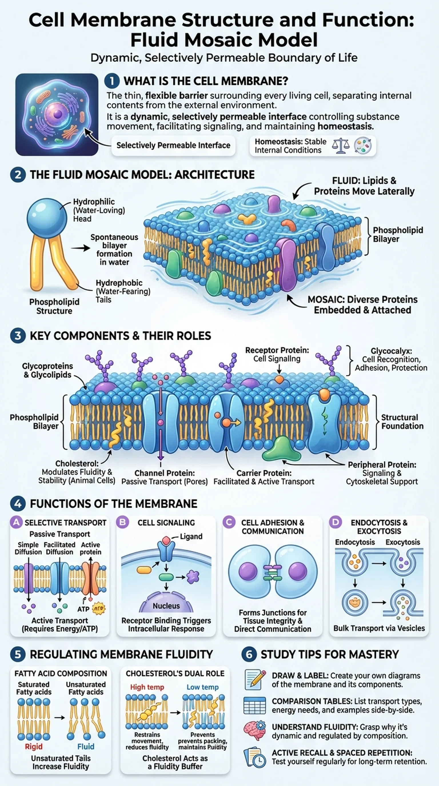
What Is the Cell Membrane?

The cell membrane, also known as the plasma membrane, is the thin, flexible barrier that surrounds every living cell and separates its internal contents from the external environment. This structure is far more than a passive boundary; it is a dynamic, selectively permeable interface that controls the movement of substances into and out of the cell, facilitates cell signaling, and maintains the internal conditions necessary for life. Understanding cell membrane structure is foundational to biology, biochemistry, and medicine.

The cell membrane is composed primarily of lipids, proteins, and carbohydrates arranged in a specific architecture described by the fluid mosaic model. This model, proposed by Singer and Nicolson in 1972, describes the membrane as a two-dimensional fluid in which lipid and protein molecules diffuse laterally. The term "mosaic" refers to the diverse collection of proteins embedded within and attached to the lipid layer, creating a patchwork of molecular components with specialized functions.

The importance of the cell membrane cannot be overstated. Without it, cells could not maintain homeostasis, the stable internal environment necessary for enzymatic reactions, DNA replication, and protein synthesis. The membrane's selective permeability ensures that essential nutrients enter the cell while waste products and potentially harmful substances are kept out or expelled. Every process in cell biology, from energy metabolism to immune response, depends on the integrity and functionality of this remarkable structure. Diseases such as cystic fibrosis and certain cancers are directly linked to defects in cell membrane proteins, underscoring the clinical relevance of understanding how the membrane is built and how it works.

Key Terms

Cell Membrane

The selectively permeable lipid bilayer that encloses the cell and regulates the transport of substances between the cell's interior and exterior.

Plasma Membrane

Another term for the cell membrane; the outermost boundary of animal cells.

Selective Permeability

The property of the cell membrane that allows certain molecules to pass through while restricting others.

Homeostasis

The maintenance of stable internal conditions within a cell or organism despite changes in the external environment.

The Fluid Mosaic Model: Architecture of the Membrane

The fluid mosaic model is the currently accepted description of cell membrane structure. It portrays the membrane as a dynamic, two-dimensional fluid composed of a lipid bilayer interspersed with a diverse array of proteins. The "fluid" component refers to the ability of individual lipid and protein molecules to move laterally within the plane of the membrane, while "mosaic" describes the varied pattern of proteins dotting the membrane surface like tiles in a mosaic artwork.

At the core of the fluid mosaic model is the phospholipid bilayer. Phospholipids are amphipathic molecules, meaning they have both a hydrophilic (water-loving) head and two hydrophobic (water-fearing) fatty acid tails. In an aqueous environment, phospholipids spontaneously arrange themselves into a bilayer with the hydrophilic heads facing outward toward the water and the hydrophobic tails tucked inward, away from water. This arrangement creates a stable yet flexible barrier that is inherently resistant to the passage of water-soluble molecules and ions.

Embedded within and associated with the phospholipid bilayer are numerous proteins that carry out the membrane's diverse functions. Integral proteins, also called transmembrane proteins, span the entire thickness of the membrane and often serve as channels, carriers, or receptors. Peripheral proteins are attached to the inner or outer surface of the membrane and typically function in cell signaling or as structural supports for the cytoskeleton. Glycoproteins and glycolipids, which have carbohydrate chains attached, extend from the outer surface and play roles in cell recognition and immune function. The fluid mosaic model thus reveals the cell membrane as a complex, highly organized structure whose fluidity is essential for processes such as membrane fusion, cell division, and endocytosis.

Key Terms

Fluid Mosaic Model

The model describing the cell membrane as a dynamic structure composed of a fluid phospholipid bilayer with a mosaic of embedded and associated proteins.

Phospholipid Bilayer

The double layer of phospholipid molecules forming the core structure of the cell membrane, with hydrophilic heads facing outward and hydrophobic tails facing inward.

Amphipathic

Having both hydrophilic and hydrophobic regions, a property that allows phospholipids to form bilayers in aqueous environments.

Integral Protein

A protein embedded in the phospholipid bilayer, often spanning the entire membrane and serving as a channel, carrier, or receptor.

Peripheral Protein

A protein loosely attached to the inner or outer surface of the cell membrane, involved in signaling or cytoskeletal support.

Components of the Cell Membrane in Detail

The cell membrane structure is composed of several molecular components, each contributing to the membrane's overall properties and functions. The phospholipid bilayer provides the structural foundation, but cholesterol, proteins, and carbohydrates add critical functionality and modulate membrane behavior.

Cholesterol molecules are interspersed among the phospholipids of animal cell membranes. Each cholesterol molecule is positioned with its hydroxyl group near the hydrophilic heads of the phospholipids and its steroid ring and hydrocarbon tail embedded in the hydrophobic core. Cholesterol has a dual effect on membrane fluidity: at high temperatures, it restrains phospholipid movement and reduces fluidity, while at low temperatures, it prevents the fatty acid tails from packing tightly and crystallizing, thereby maintaining fluidity. This buffering function is essential for keeping the cell membrane functional across a range of physiological temperatures.

Transport proteins are among the most important components of cell membrane structure. Channel proteins form pores that allow specific ions or small molecules to pass through the membrane passively, following their concentration gradient. Carrier proteins undergo conformational changes to shuttle molecules across the membrane, and some require energy in the form of ATP to move substances against their gradient. Receptor proteins on the membrane surface bind specific signaling molecules such as hormones and neurotransmitters, triggering intracellular responses through signal transduction cascades.

Carbohydrates are found exclusively on the extracellular surface of the cell membrane, attached to lipids (glycolipids) or proteins (glycoproteins). This carbohydrate-rich layer, called the glycocalyx, functions in cell-cell recognition, adhesion, and protection. The glycocalyx allows immune cells to distinguish self from non-self, which is critical for immune surveillance and transplant compatibility. Together, these molecular components create the intricate and versatile architecture described by the fluid mosaic model.

Key Terms

Cholesterol

A steroid lipid embedded in the cell membrane that modulates fluidity by preventing extremes of rigidity and fluidity.

Channel Protein

A transmembrane protein that forms a hydrophilic pore, allowing specific ions or molecules to pass through the membrane.

Glycocalyx

The carbohydrate-rich layer on the outer surface of the cell membrane formed by glycoproteins and glycolipids, involved in cell recognition.

Signal Transduction

The process by which a signal from outside the cell is transmitted to the interior, often initiated by receptor proteins in the cell membrane.

Functions of the Cell Membrane

The cell membrane performs a remarkable range of functions that are essential for cell survival, communication, and organization. These functions arise directly from the membrane's unique structure, as described by the fluid mosaic model, and are made possible by the interplay of its lipid, protein, and carbohydrate components.

The most fundamental function of the cell membrane is selective transport. The phospholipid bilayer is inherently impermeable to most polar molecules, ions, and large macromolecules. Small nonpolar molecules such as oxygen and carbon dioxide can diffuse freely across the membrane, but the transport of glucose, amino acids, and ions requires specialized proteins. Passive transport mechanisms, including simple diffusion, facilitated diffusion, and osmosis, move substances down their concentration gradients without energy expenditure. Active transport mechanisms use ATP or the energy stored in electrochemical gradients to move substances against their concentration gradients, maintaining the precise ion concentrations that cells need for signaling and metabolism.

Cell signaling is another critical function. Receptor proteins on the cell membrane surface bind extracellular ligands such as hormones, growth factors, and neurotransmitters. This binding event triggers conformational changes in the receptor that activate intracellular signaling pathways, ultimately altering gene expression, metabolism, or cell behavior. Without functional membrane receptors, cells cannot respond to their environment, which can lead to uncontrolled growth (cancer) or immune deficiency.

The cell membrane also facilitates cell adhesion and communication. Membrane proteins form junctions between adjacent cells, including tight junctions, gap junctions, and desmosomes, which hold tissues together and allow direct communication between neighboring cells. Endocytosis and exocytosis, processes in which the membrane engulfs or releases materials in vesicles, enable the cell to take in nutrients, secrete proteins, and recycle membrane components. These diverse functions illustrate why the cell membrane structure is central to understanding all of biology.

Key Terms

Active Transport

The movement of molecules across the cell membrane against their concentration gradient, requiring energy input, typically from ATP.

Facilitated Diffusion

The passive transport of molecules across the membrane through specific protein channels or carriers, without energy expenditure.

Endocytosis

The process by which the cell membrane engulfs extracellular material, forming a vesicle that is brought into the cell.

Exocytosis

The process by which vesicles fuse with the cell membrane to release their contents outside the cell.

Membrane Fluidity and Its Regulation

Membrane fluidity is a critical property of the cell membrane that affects nearly every aspect of membrane function, from transport and signaling to cell division and immune response. The fluid mosaic model emphasizes that the membrane is not a rigid structure but a dynamic, viscous fluid in which lipids and proteins are constantly in motion. The degree of fluidity must be carefully regulated to ensure optimal membrane performance under varying environmental conditions.

Several factors influence the fluidity of the phospholipid bilayer. The length and saturation of fatty acid tails play a major role. Shorter fatty acid chains have fewer van der Waals interactions with neighboring chains and increase fluidity. Unsaturated fatty acids, which contain one or more double bonds, introduce kinks in the hydrocarbon tails that prevent tight packing and further enhance fluidity. In contrast, long, saturated fatty acid tails pack closely together, reducing fluidity and making the membrane more rigid.

Cholesterol is the primary regulator of membrane fluidity in animal cells. As discussed earlier, cholesterol inserts itself between phospholipids and acts as a fluidity buffer: at high temperatures it restricts phospholipid movement, and at low temperatures it disrupts orderly packing. This dual role allows the cell membrane to maintain a consistent viscosity, a property sometimes called homeoviscous adaptation. Organisms that live in cold environments, such as deep-sea fish, tend to have membranes with a higher proportion of unsaturated fatty acids to prevent the membrane from becoming too rigid.

The regulation of membrane fluidity has direct implications for cell membrane structure and function. If the membrane becomes too rigid, transport proteins cannot undergo the conformational changes needed for active transport, and receptor-mediated signaling is impaired. If the membrane becomes too fluid, it loses its barrier function and structural integrity. Understanding these dynamics is essential for comprehending drug interactions with membranes, the mechanism of anesthetics, and diseases that involve membrane dysfunction.

Key Terms

Membrane Fluidity

The viscosity of the lipid bilayer, determined by fatty acid composition and cholesterol content, affecting protein mobility and membrane function.

Unsaturated Fatty Acid

A fatty acid with one or more double bonds in its hydrocarbon tail, creating kinks that increase membrane fluidity.

Saturated Fatty Acid

A fatty acid with no double bonds, allowing tight packing that decreases membrane fluidity.

Homeoviscous Adaptation

The adjustment of membrane lipid composition to maintain optimal fluidity in response to temperature changes.

Study Tips for Mastering Cell Membrane Biology

The cell membrane is one of the most frequently tested topics in biology courses, appearing on exams from AP Biology to the MCAT and medical school histology courses. Mastering cell membrane structure and the fluid mosaic model requires a systematic approach that combines visual learning with active problem-solving.

First, build a layered mental model of the membrane. Start with the phospholipid bilayer as the foundation, then add cholesterol, integral proteins, peripheral proteins, and the glycocalyx one component at a time. For each component, learn its structure, its position in the membrane, and its function. Drawing the cell membrane by hand, labeling each component, is one of the most effective study techniques available. Annotate your diagram with arrows showing the direction of transport for different molecules.

Second, create comparison tables for the different types of membrane transport. List simple diffusion, facilitated diffusion, osmosis, active transport, endocytosis, and exocytosis side by side with columns for energy requirement, direction of movement, type of molecule transported, and examples. This organized approach helps you quickly recall distinctions during exams. Third, understand the fluid mosaic model not just as a static description but as a dynamic concept. Ask yourself: why is fluidity important? What happens to cell function if the membrane becomes too rigid or too fluid? How does cholesterol regulate this balance?

Finally, test yourself using active recall and spaced repetition. Platforms like LectureScribe can convert your lecture notes into flashcards and practice questions focused on cell membrane structure, the phospholipid bilayer, and membrane transport mechanisms. Repeated self-testing is significantly more effective than passive review for long-term retention. Make sure you can explain the fluid mosaic model in your own words and apply your knowledge to novel scenarios, such as predicting the effects of a mutation in a specific membrane protein.

Key Terms

Active Recall

A study technique that involves actively retrieving information from memory rather than passively reviewing notes.

Spaced Repetition

A learning strategy that involves reviewing material at increasing intervals to strengthen long-term memory retention.

MCAT

The Medical College Admission Test; a standardized exam required for admission to medical schools in the United States and Canada.

Frequently Asked Questions

What is the cell membrane and what does it do?

The cell membrane is the selectively permeable barrier surrounding every cell. It controls the movement of substances in and out of the cell, facilitates cell signaling, maintains homeostasis, and provides structural support. Its structure is described by the fluid mosaic model.

What is the fluid mosaic model?

The fluid mosaic model describes the cell membrane as a dynamic structure composed of a fluid phospholipid bilayer interspersed with a mosaic of proteins, cholesterol, and carbohydrates. Lipids and proteins can move laterally within the membrane, giving it flexibility and versatility.

What is the phospholipid bilayer?

The phospholipid bilayer is the double layer of phospholipid molecules that forms the core structure of the cell membrane. The hydrophilic heads face the aqueous environment on both sides, while the hydrophobic tails face inward, creating a barrier to water-soluble substances.

What is the role of cholesterol in the cell membrane?

Cholesterol regulates membrane fluidity by inserting between phospholipids. At high temperatures, it reduces fluidity by restraining phospholipid movement. At low temperatures, it prevents the membrane from becoming too rigid by disrupting tight packing of fatty acid tails.

What is the difference between integral and peripheral proteins?

Integral proteins are embedded in or span the entire phospholipid bilayer, often functioning as channels, carriers, or receptors. Peripheral proteins are loosely attached to the inner or outer membrane surface and typically function in signaling or cytoskeletal support.

How does the cell membrane structure relate to its function?

The phospholipid bilayer provides a selective barrier, integral proteins enable transport and signaling, cholesterol regulates fluidity, and glycoproteins allow cell recognition. This diverse architecture, described by the fluid mosaic model, enables the membrane to perform its many functions.

Why is membrane fluidity important?

Membrane fluidity is essential because it allows transport proteins to change shape, enables membrane fusion during endocytosis and exocytosis, permits cell growth and division, and ensures proper receptor function. Without appropriate fluidity, cell membrane function is severely impaired.

Study Biology Smarter

Upload your lecture notes and get AI-generated flashcards, quizzes, infographics, and study guides tailored to your curriculum.

Try LectureScribe Free

Create Study Materials

Transform your Biology lectures into flashcards, quizzes, and visual study guides with AI.

  • AI-generated flashcards & quizzes
  • Visual infographics from notes
  • Interactive study shorts
Start Studying Free

Quick Facts

SubjectBiology
Word Count2,500+
Slides5
InfographicIncluded
AuthorLectureScribe

Master Biology with LectureScribe

Upload your lecture notes and get AI-powered flashcards, quizzes, infographics, and study guides in minutes.