LectureScribe Logo
lecturescribe.io

LOCK IN VALENTINE PRICING BEFORE SALE ENDS!

Use code — New joiners get 40% OFF on annual plans

Plans will have higher prices after this sale ends

0days
:
0hours
:
0minutes
:
0seconds
Anatomy2,500+ words5 slides
🫁

Digestive System: Organs, Functions & Enzyme Guide

Learn the digestive system organs, functions, and key digestive enzymes. Understand the complete digestion process through the GI tract from mouth to large intestine.

L
LectureScribeAI-Powered Study Platform

Study Infographic

Complete digestive system diagram illustrating the GI tract organs, accessory digestive organs, and key digestive enzymes involved in the digestion process.

Download
Digestive system diagram showing all digestive organs of the GI tract with digestive enzymes and their functions labeled

Interactive Study Short

Swipe through 5 slides about Digestive System: Organs, Functions & Enzyme Guide

1 / 5

Overview of the Digestive System

The digestive system is a complex organ system responsible for breaking down food into nutrients that the body can absorb and use for energy, growth, and cellular repair. Extending from the mouth to the anus, the digestive system encompasses a continuous muscular tube known as the gastrointestinal tract, or GI tract, along with several accessory organs that secrete digestive enzymes and other substances essential for the digestion process.

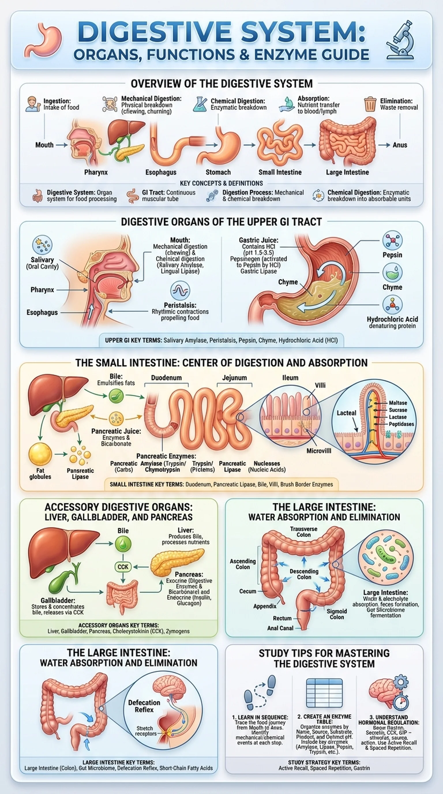
The primary functions of the digestive system include ingestion, mechanical digestion, chemical digestion, absorption, and elimination. Ingestion is the intake of food through the mouth. Mechanical digestion involves the physical breakdown of food into smaller pieces through chewing, churning, and segmentation. Chemical digestion uses digestive enzymes and other chemicals to break down macromolecules such as proteins, carbohydrates, and lipids into their absorbable subunits. Absorption is the transfer of nutrients from the GI tract lumen into the bloodstream and lymphatic system. Finally, elimination is the removal of indigestible waste from the body as feces.

Understanding the digestive system is foundational for students of anatomy, physiology, nutrition, and medicine. The digestive organs work in a coordinated sequence, each contributing specific mechanical or chemical actions to the overall digestion process. Disruptions at any point along the GI tract can lead to clinical conditions ranging from gastroesophageal reflux disease and peptic ulcers to malabsorption syndromes and inflammatory bowel disease. By studying the structure and function of each digestive organ, students gain the knowledge needed to understand both normal physiology and the pathophysiology of gastrointestinal disorders.

Key Terms

Digestive System

The organ system responsible for the ingestion, digestion, absorption, and elimination of food and nutrients.

GI Tract

The gastrointestinal tract; the continuous muscular tube from the mouth to the anus through which food passes during digestion.

Digestion Process

The complete sequence of mechanical and chemical events that break down food into absorbable nutrients within the digestive system.

Chemical Digestion

The enzymatic breakdown of macromolecules into smaller molecules that can be absorbed across the intestinal lining.

Digestive Organs of the Upper GI Tract

The digestive organs of the upper gastrointestinal tract include the mouth, pharynx, esophagus, and stomach. Each of these organs plays a distinct role in the early stages of the digestion process, preparing food for the extensive chemical digestion and absorption that occurs in the small intestine.

The mouth, or oral cavity, is where the digestion process begins. Mechanical digestion starts with mastication (chewing), which breaks food into smaller particles and increases the surface area available for enzymatic action. The salivary glands secrete saliva, which contains the digestive enzyme salivary amylase (also called ptyalin). Salivary amylase initiates the chemical digestion of starch, breaking it into maltose and dextrins. Saliva also contains lingual lipase, which begins the digestion of fats, and mucus, which lubricates the food bolus for swallowing.

The pharynx and esophagus serve primarily as conduits for transporting the food bolus from the mouth to the stomach. Swallowing (deglutition) is a coordinated reflex that propels food through the pharynx and into the esophagus, where rhythmic smooth muscle contractions called peristalsis move the bolus toward the stomach. The lower esophageal sphincter prevents the reflux of acidic stomach contents back into the esophagus.

The stomach is a J-shaped muscular organ that serves as a temporary storage reservoir and a site of significant mechanical and chemical digestion. The muscular walls of the stomach churn food, mixing it with gastric juice to produce a semi-liquid mixture called chyme. Gastric juice contains hydrochloric acid (HCl), which creates a highly acidic environment (pH 1.5-3.5) that denatures proteins and activates the digestive enzyme pepsinogen into its active form, pepsin. Pepsin is a protease that begins breaking down proteins into smaller peptide fragments. The stomach also secretes gastric lipase, which contributes to fat digestion, and intrinsic factor, which is essential for vitamin B12 absorption in the small intestine. These digestive organs of the upper GI tract collectively transform food from a solid mass into chyme, ready for further processing downstream.

Key Terms

Salivary Amylase

A digestive enzyme secreted by the salivary glands that initiates the chemical digestion of starch in the mouth.

Peristalsis

Rhythmic waves of smooth muscle contraction that propel food along the GI tract.

Pepsin

A gastric protease enzyme activated by hydrochloric acid that breaks down proteins into smaller peptide fragments in the stomach.

Chyme

The semi-liquid mixture of partially digested food and gastric secretions produced in the stomach.

Hydrochloric Acid (HCl)

A strong acid secreted by parietal cells in the stomach that denatures proteins, activates pepsinogen, and kills pathogens.

The Small Intestine: Center of Digestion and Absorption

The small intestine is the longest segment of the GI tract, measuring approximately 6 meters in length, and it is the primary site of both chemical digestion and nutrient absorption. The small intestine is divided into three anatomical regions: the duodenum, jejunum, and ileum. Each region contributes to the digestion process in specific ways, and together they ensure that nearly all nutrients from ingested food are broken down and absorbed into the body.

The duodenum is the first and shortest segment of the small intestine, receiving chyme from the stomach along with secretions from the pancreas and liver. Pancreatic juice, delivered via the pancreatic duct, contains a powerful cocktail of digestive enzymes including pancreatic amylase (which continues starch digestion), trypsin and chymotrypsin (which break down proteins), pancreatic lipase (which digests fats), and nucleases (which digest nucleic acids). Pancreatic juice also contains bicarbonate, which neutralizes the acidic chyme and creates the slightly alkaline environment (pH 7-8) optimal for intestinal enzyme activity. Bile, produced by the liver and stored in the gallbladder, is released into the duodenum to emulsify fats, breaking large lipid droplets into smaller micelles that pancreatic lipase can act upon more efficiently.

The jejunum is the middle segment and the most active site of nutrient absorption. Its mucosal lining is characterized by circular folds (plicae circulares), villi, and microvilli, which collectively increase the absorptive surface area by approximately 600-fold. Brush border digestive enzymes, such as maltase, sucrase, lactase, and peptidases, complete the final stages of carbohydrate and protein digestion at the surface of the epithelial cells. The products of digestion, including monosaccharides, amino acids, fatty acids, and monoglycerides, are absorbed through the epithelium and enter either the capillary network (for water-soluble nutrients) or the lacteals (for lipid-soluble nutrients).

The ileum is the final segment of the small intestine and is specialized for the absorption of vitamin B12, bile salts, and any remaining nutrients not absorbed in the jejunum. The ileocecal valve at the junction of the ileum and large intestine regulates the passage of material into the colon and prevents backflow. Together, the three regions of the small intestine represent the most critical digestive organs for completing the digestion process and extracting nutrition from food.

Key Terms

Duodenum

The first segment of the small intestine where chyme mixes with pancreatic juice and bile for extensive chemical digestion.

Pancreatic Lipase

A digestive enzyme secreted by the pancreas that breaks down triglycerides into fatty acids and monoglycerides.

Bile

A greenish-yellow fluid produced by the liver that emulsifies fats in the duodenum, facilitating their digestion by lipase.

Villi

Finger-like projections of the small intestinal mucosa that increase the surface area for nutrient absorption.

Brush Border Enzymes

Digestive enzymes located on the microvilli of intestinal epithelial cells that complete the final stages of carbohydrate and protein digestion.

Accessory Digestive Organs: Liver, Gallbladder, and Pancreas

The accessory digestive organs, including the liver, gallbladder, and pancreas, do not directly contact food as it moves through the GI tract, but they produce and deliver essential secretions that are indispensable for the digestion process. Without these accessory organs, the chemical digestion of fats, proteins, and carbohydrates in the small intestine would be severely impaired.

The liver is the largest internal organ in the body and performs hundreds of metabolic functions. In the context of the digestive system, its primary role is the production of bile. Bile contains bile salts, cholesterol, phospholipids, bilirubin, and water. Bile salts are the functionally important component for digestion: they act as emulsifying agents that break large fat globules into smaller droplets, dramatically increasing the surface area available for pancreatic lipase to act upon. The liver also plays critical roles in nutrient processing, detoxification, and storage. After absorption, nutrient-rich blood from the GI tract travels via the hepatic portal vein to the liver, where it is processed before entering the general circulation.

The gallbladder is a small, pear-shaped organ located beneath the liver that stores and concentrates bile between meals. When fatty chyme enters the duodenum, the hormone cholecystokinin (CCK) is released by enteroendocrine cells in the intestinal wall. CCK stimulates the gallbladder to contract and release concentrated bile into the common bile duct, which empties into the duodenum. Gallstones, formed from crystallized cholesterol or bilirubin, can obstruct the bile duct and impair fat digestion.

The pancreas is both an exocrine and endocrine gland. Its exocrine function, relevant to the digestive system, involves the production and secretion of pancreatic juice containing digestive enzymes and bicarbonate. The pancreas produces the full spectrum of enzymes needed to digest carbohydrates (pancreatic amylase), proteins (trypsin, chymotrypsin, carboxypeptidase, elastase), fats (pancreatic lipase, colipase), and nucleic acids (nucleases). Many of these digestive enzymes are secreted as inactive zymogens (such as trypsinogen) to prevent self-digestion, and they are activated only upon reaching the duodenum. The hormone secretin stimulates the pancreas to release bicarbonate, while CCK stimulates enzyme secretion. Together, the liver, gallbladder, and pancreas form a triad of accessory digestive organs that are essential for efficient nutrient breakdown.

Key Terms

Liver

The largest internal organ; produces bile for fat emulsification and processes absorbed nutrients via the hepatic portal system.

Gallbladder

A small organ that stores and concentrates bile, releasing it into the duodenum in response to cholecystokinin (CCK).

Pancreas

A dual-function organ that secretes digestive enzymes and bicarbonate (exocrine) and hormones like insulin (endocrine).

Cholecystokinin (CCK)

A hormone released by the small intestine that stimulates gallbladder contraction and pancreatic enzyme secretion.

Zymogens

Inactive enzyme precursors (e.g., trypsinogen, pepsinogen) that are activated in the GI tract to prevent premature self-digestion.

The Large Intestine: Water Absorption and Elimination

The large intestine, also called the colon, is the final major segment of the GI tract and plays a crucial role in water absorption, electrolyte balance, and the formation and elimination of feces. While the large intestine does not produce significant digestive enzymes or participate in major chemical digestion, it is an essential component of the digestive system that completes the digestion process.

Anatomically, the large intestine is approximately 1.5 meters long and is divided into several regions: the cecum, ascending colon, transverse colon, descending colon, sigmoid colon, rectum, and anal canal. The cecum is a pouch-like structure that receives material from the ileum through the ileocecal valve. The appendix, a small, finger-like projection attached to the cecum, is thought to play a role in immune function and as a reservoir for beneficial gut bacteria.

The primary function of the large intestine is the absorption of water and electrolytes (sodium, potassium, chloride) from the remaining indigestible material. Approximately 1.5 liters of fluid enters the large intestine daily, and about 90% of this water is reabsorbed, transforming liquid chyme into semi-solid feces. The colon also absorbs vitamins produced by the gut microbiota, particularly vitamin K and certain B vitamins. The trillions of bacteria residing in the large intestine, collectively called the gut microbiome, ferment undigested carbohydrates and fiber, producing short-chain fatty acids that nourish the colonocytes and contribute to overall health.

Mass movements, a type of strong peristaltic contraction unique to the large intestine, propel feces toward the rectum. When the rectum is distended with feces, stretch receptors trigger the defecation reflex, a coordinated response involving relaxation of the internal anal sphincter, voluntary relaxation of the external anal sphincter, and contraction of the abdominal muscles. Disorders of the large intestine, including irritable bowel syndrome, ulcerative colitis, Crohn's disease, and colorectal cancer, are among the most common gastrointestinal conditions encountered in clinical practice. A thorough understanding of large intestine anatomy and function is therefore essential for any student studying the digestive organs and the digestive system as a whole.

Key Terms

Large Intestine (Colon)

The final major segment of the GI tract responsible for water absorption, electrolyte balance, and feces formation.

Gut Microbiome

The diverse community of trillions of microorganisms residing in the large intestine that aid in fermentation, vitamin production, and immune function.

Defecation Reflex

A coordinated reflex triggered by rectal distension that results in the elimination of feces from the body.

Short-Chain Fatty Acids

Metabolic products of bacterial fermentation in the colon (e.g., butyrate, propionate, acetate) that nourish colonocytes and support gut health.

Study Tips for Mastering the Digestive System

The digestive system is one of the most content-rich topics in anatomy and physiology courses, and it is heavily tested on exams such as the USMLE, MCAT, and nursing board examinations. Success requires a systematic approach that integrates structural anatomy with functional physiology and clinical applications.

First, learn the digestive organs in sequence by following the path of a meal through the GI tract. Start at the mouth and trace the journey through the pharynx, esophagus, stomach, duodenum, jejunum, ileum, cecum, ascending colon, transverse colon, descending colon, sigmoid colon, rectum, and anal canal. At each stop, identify the key mechanical and chemical events, the digestive enzymes involved, and the nutrients absorbed. This journey-based approach creates a logical narrative that is easier to remember than isolated facts.

Second, create a comprehensive digestive enzymes table. Organize your table with columns for enzyme name, source organ, substrate (what it digests), product (what it produces), and optimal pH. Include salivary amylase, lingual lipase, pepsin, pancreatic amylase, trypsin, chymotrypsin, pancreatic lipase, maltase, sucrase, lactase, and peptidases. This table is one of the highest-yield study aids for the digestive system, as enzyme-related questions appear frequently on standardized exams.

Third, understand the hormonal regulation of the digestion process. The four major GI hormones to know are gastrin (stimulates gastric acid secretion), secretin (stimulates bicarbonate release from the pancreas), cholecystokinin (stimulates gallbladder contraction and pancreatic enzyme secretion), and gastric inhibitory peptide (inhibits gastric acid secretion and stimulates insulin release). Knowing the stimulus, source, and action of each hormone will help you answer regulation-based questions with confidence.

Finally, use active recall and spaced repetition to solidify your knowledge of the digestive system and its many components. Platforms like LectureScribe can generate flashcards, slide decks, and practice questions directly from your digestive system lecture notes, helping you review the digestive organs, digestive enzymes, and the complete digestion process on a regular schedule for long-term retention.

Key Terms

Active Recall

A study technique in which learners actively retrieve information from memory rather than passively reviewing notes.

Spaced Repetition

A learning strategy that involves reviewing material at increasing intervals to strengthen long-term memory retention.

Gastrin

A GI hormone secreted by G cells in the stomach that stimulates hydrochloric acid production and gastric motility.

Frequently Asked Questions

What are the main organs of the digestive system?

The digestive organs include the mouth, pharynx, esophagus, stomach, small intestine (duodenum, jejunum, ileum), and large intestine (colon, rectum, anal canal). Accessory organs include the liver, gallbladder, and pancreas. Together these organs form the complete GI tract and digestive system.

What are the most important digestive enzymes?

Key digestive enzymes include salivary amylase (starch digestion in the mouth), pepsin (protein digestion in the stomach), pancreatic amylase, trypsin, chymotrypsin, and pancreatic lipase (carbohydrate, protein, and fat digestion in the small intestine), and brush border enzymes like maltase, sucrase, and lactase.

What is the GI tract?

The GI tract (gastrointestinal tract) is the continuous muscular tube that extends from the mouth to the anus. It includes the oral cavity, pharynx, esophagus, stomach, small intestine, and large intestine. The GI tract is where the digestion process physically takes place.

What is the digestion process?

The digestion process is the complete sequence of mechanical and chemical events that break food down into absorbable nutrients. It begins with ingestion and mechanical digestion in the mouth, continues with chemical digestion by digestive enzymes in the stomach and small intestine, and concludes with absorption and elimination.

Where does most nutrient absorption occur in the digestive system?

Most nutrient absorption occurs in the small intestine, particularly the jejunum. The small intestine's enormous surface area, created by circular folds, villi, and microvilli, allows for efficient absorption of monosaccharides, amino acids, fatty acids, vitamins, and minerals.

What role does the pancreas play in digestion?

The pancreas produces pancreatic juice containing digestive enzymes (amylase, lipase, trypsin, chymotrypsin) and bicarbonate. These secretions are delivered to the duodenum where they perform the majority of chemical digestion of carbohydrates, proteins, and fats in the GI tract.

How does bile help in fat digestion?

Bile, produced by the liver and stored in the gallbladder, emulsifies fats by breaking large lipid droplets into smaller micelles. This dramatically increases the surface area available for pancreatic lipase, making fat digestion much more efficient. Without bile, fat digestion and absorption would be severely impaired.

Study Anatomy Smarter

Upload your lecture notes and get AI-generated flashcards, quizzes, infographics, and study guides tailored to your curriculum.

Try LectureScribe Free

Master Anatomy with LectureScribe

Upload your lecture notes and get AI-powered flashcards, quizzes, infographics, and study guides in minutes.