LectureScribe Logo
lecturescribe.io

LOCK IN VALENTINE PRICING BEFORE SALE ENDS!

Use code — New joiners get 40% OFF on annual plans

Plans will have higher prices after this sale ends

0days
:
0hours
:
0minutes
:
0seconds
Anatomy2,500+ words5 slides
🫁

Respiratory System: Gas Exchange and Breathing Mechanics

Learn the respiratory system, gas exchange in the alveoli, and breathing mechanics. Understand pulmonary ventilation, oxygen transport, and the anatomy of respiration.

L
LectureScribeAI-Powered Study Platform

Study Infographic

Complete respiratory system diagram illustrating airway anatomy, alveoli structure, gas exchange at the respiratory membrane, and the mechanics of pulmonary ventilation.

Download
Respiratory system diagram showing the airways, alveoli, gas exchange process, and breathing mechanics of pulmonary ventilation

Interactive Study Short

Swipe through 5 slides about Respiratory System: Gas Exchange and Breathing Mechanics

1 / 5

Overview of the Respiratory System

The respiratory system is the organ system responsible for the exchange of gases between the body and the external environment. Its primary function is to deliver oxygen to the blood and remove carbon dioxide, a metabolic waste product, from the body. The respiratory system works in close partnership with the cardiovascular system to ensure that every cell in the body receives the oxygen it needs for aerobic metabolism and that carbon dioxide is efficiently eliminated.

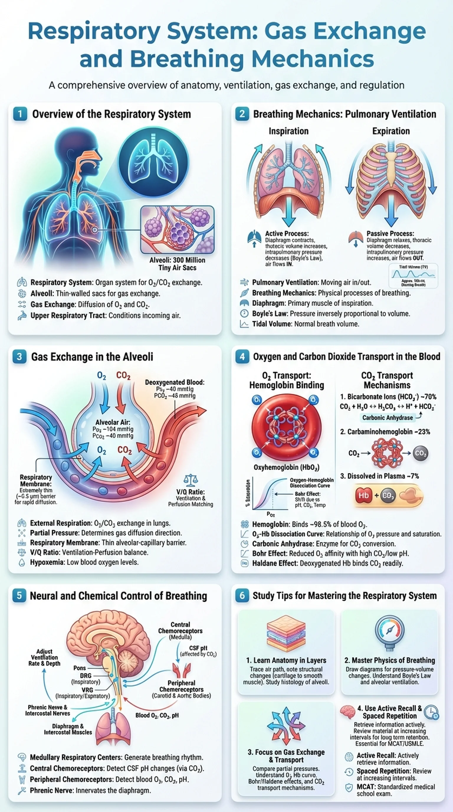
Anatomically, the respiratory system is divided into the upper respiratory tract and the lower respiratory tract. The upper respiratory tract includes the nose, nasal cavity, pharynx, and larynx. These structures warm, humidify, and filter the incoming air before it reaches the lungs. The lower respiratory tract includes the trachea, bronchi, bronchioles, and lungs, where the actual process of gas exchange occurs. The lungs contain approximately 300 million alveoli, tiny air sacs with extremely thin walls that provide an enormous surface area (approximately 70 square meters) for the diffusion of oxygen and carbon dioxide.

Understanding the respiratory system is essential for students of anatomy, physiology, and medicine. Respiratory diseases, including asthma, chronic obstructive pulmonary disease (COPD), pneumonia, and pulmonary embolism, are among the leading causes of morbidity and mortality worldwide. A solid grasp of respiratory anatomy, breathing mechanics, and the principles of gas exchange enables students to understand how these diseases disrupt normal function and how treatments work to restore it. In this article, we will explore the anatomy of the respiratory system, the mechanics of pulmonary ventilation, the process of gas exchange in the alveoli, and the transport of oxygen and carbon dioxide in the blood.

Key Terms

Respiratory System

The organ system responsible for gas exchange, delivering oxygen to the blood and removing carbon dioxide from the body.

Alveoli

Tiny, thin-walled air sacs in the lungs where gas exchange between air and blood occurs across the respiratory membrane.

Gas Exchange

The diffusion of oxygen from the alveoli into the pulmonary capillaries and carbon dioxide from the capillaries into the alveoli.

Upper Respiratory Tract

The portion of the respiratory system that includes the nose, nasal cavity, pharynx, and larynx, which condition incoming air.

Breathing Mechanics: Pulmonary Ventilation

Breathing mechanics, also known as pulmonary ventilation, refers to the physical process of moving air into and out of the lungs. Pulmonary ventilation is driven by pressure gradients created by changes in the volume of the thoracic cavity, and it consists of two phases: inspiration (inhalation) and expiration (exhalation). Understanding breathing mechanics is fundamental to appreciating how the respiratory system delivers fresh air to the alveoli for gas exchange.

Inspiration is an active process that requires muscular effort. The primary muscle of inspiration is the diaphragm, a dome-shaped skeletal muscle that separates the thoracic and abdominal cavities. When the diaphragm contracts, it flattens and moves downward, increasing the vertical dimension of the thoracic cavity. Simultaneously, the external intercostal muscles contract, pulling the ribs upward and outward, further expanding the thoracic cavity. According to Boyle's Law, as the volume of the thoracic cavity increases, the intrapulmonary pressure (pressure inside the lungs) decreases below atmospheric pressure. This negative pressure gradient causes air to flow into the lungs through the airways, filling the alveoli with fresh, oxygen-rich air.

Expiration during quiet breathing is primarily a passive process. When the diaphragm and external intercostals relax, the elastic recoil of the lungs and chest wall returns the thoracic cavity to its resting volume. As the thoracic cavity decreases in size, intrapulmonary pressure rises above atmospheric pressure, and air flows out of the lungs. During forced expiration, such as during exercise or coughing, the internal intercostal muscles and abdominal muscles actively contract to compress the thoracic cavity and expel air more forcefully.

Several lung volumes and capacities are used to quantify the effectiveness of pulmonary ventilation and breathing mechanics. Tidal volume is the amount of air moved in or out during a normal breath (approximately 500 mL). Vital capacity is the maximum volume of air that can be exhaled after a maximal inhalation. Residual volume is the air remaining in the lungs after a maximal exhalation, preventing the alveoli from collapsing. These measurements, obtained through spirometry, are essential clinical tools for assessing respiratory system function and diagnosing conditions such as restrictive and obstructive lung diseases.

Key Terms

Pulmonary Ventilation

The process of moving air into and out of the lungs through the mechanical actions of breathing; also called breathing mechanics.

Breathing Mechanics

The physical processes involving muscular contraction, pressure changes, and air flow that accomplish pulmonary ventilation.

Diaphragm

The primary muscle of inspiration; a dome-shaped skeletal muscle whose contraction increases thoracic cavity volume and draws air into the lungs.

Boyle's Law

The gas law stating that at constant temperature, the pressure of a gas is inversely proportional to its volume; explains how thoracic expansion creates airflow.

Tidal Volume

The volume of air inhaled or exhaled during a normal, resting breath, approximately 500 mL in an average adult.

Gas Exchange in the Alveoli

Gas exchange is the core function of the respiratory system and occurs at two sites in the body: the alveoli of the lungs (external respiration) and the tissues (internal respiration). External respiration, the exchange of gases between the alveoli and the pulmonary capillaries, is the focus of this section and represents the critical link between the air we breathe and the oxygen that reaches our cells.

Gas exchange in the alveoli is driven by the principle of diffusion: gases move from areas of higher partial pressure to areas of lower partial pressure across a permeable membrane. Inhaled air in the alveoli has a high partial pressure of oxygen (approximately 104 mmHg) and a low partial pressure of carbon dioxide (approximately 40 mmHg). Deoxygenated blood arriving in the pulmonary capillaries has a low partial pressure of oxygen (approximately 40 mmHg) and a high partial pressure of carbon dioxide (approximately 45 mmHg). Oxygen therefore diffuses down its concentration gradient from the alveoli into the blood, while carbon dioxide diffuses from the blood into the alveoli to be exhaled.

The efficiency of gas exchange depends on several factors. First, the respiratory membrane, consisting of the alveolar epithelium, the capillary endothelium, and their fused basement membranes, must be extremely thin (approximately 0.5 micrometers) to allow rapid diffusion. Second, the enormous surface area provided by the approximately 300 million alveoli ensures that a large volume of gas can be exchanged simultaneously. Third, the alveoli must be adequately ventilated (receiving fresh air through pulmonary ventilation) and perfused (supplied with blood flow through the pulmonary capillaries). The matching of ventilation and perfusion, known as the V/Q ratio, is a critical concept in respiratory physiology. When ventilation and perfusion are well matched, gas exchange is optimized. Conditions such as pulmonary embolism (which reduces perfusion) or airway obstruction (which reduces ventilation) disrupt the V/Q ratio and impair gas exchange, leading to hypoxemia (low blood oxygen) and potentially respiratory failure.

Key Terms

External Respiration

The exchange of oxygen and carbon dioxide between the alveoli and the pulmonary capillaries in the lungs.

Partial Pressure

The pressure exerted by a single gas in a mixture of gases; determines the direction of gas diffusion during gas exchange.

Respiratory Membrane

The thin barrier between alveolar air and capillary blood, consisting of the alveolar and capillary walls and their fused basement membranes.

V/Q Ratio

The ratio of alveolar ventilation to pulmonary capillary perfusion; optimal matching is essential for efficient gas exchange.

Hypoxemia

Abnormally low oxygen levels in the arterial blood, often caused by impaired gas exchange or ventilation-perfusion mismatch.

Oxygen and Carbon Dioxide Transport in the Blood

Once gas exchange has occurred in the alveoli, oxygen must be transported from the lungs to the tissues, and carbon dioxide must be carried from the tissues back to the lungs. The respiratory system depends on the circulatory system to accomplish this transport, and the blood uses several mechanisms to carry these gases efficiently.

Oxygen transport occurs primarily through binding to hemoglobin, a protein found within red blood cells. Each hemoglobin molecule contains four heme groups, each of which can bind one molecule of oxygen, giving each hemoglobin the capacity to carry four oxygen molecules. Approximately 98.5% of oxygen in the blood is transported bound to hemoglobin as oxyhemoglobin, while only 1.5% is dissolved directly in the plasma. The oxygen-hemoglobin dissociation curve describes the relationship between the partial pressure of oxygen and the percentage of hemoglobin saturated with oxygen. This sigmoidal curve reflects the cooperative binding of oxygen: as one heme group binds oxygen, the remaining groups bind with increasing affinity. Factors such as pH, temperature, carbon dioxide levels, and 2,3-bisphosphoglycerate (2,3-BPG) shift the curve, modulating oxygen release to match tissue demands.

Carbon dioxide transport from the tissues to the lungs occurs via three mechanisms. Approximately 70% of carbon dioxide is transported as bicarbonate ions (HCO3-), formed when CO2 reacts with water in red blood cells in a reaction catalyzed by the enzyme carbonic anhydrase. About 23% of carbon dioxide binds to hemoglobin (at a different site than oxygen) to form carbaminohemoglobin. The remaining 7% is dissolved directly in the plasma. At the alveoli, these reactions reverse: bicarbonate is converted back to CO2, carbaminohemoglobin releases CO2, and dissolved CO2 diffuses across the respiratory membrane into the alveoli for exhalation.

The interdependence of oxygen and carbon dioxide transport is illustrated by two important physiological phenomena. The Bohr effect describes how increased CO2 and decreased pH in the tissues promote oxygen release from hemoglobin, enhancing oxygen delivery where it is needed most. The Haldane effect describes how deoxygenated hemoglobin has a greater affinity for CO2 and hydrogen ions, facilitating carbon dioxide pickup in the tissues. Together, the Bohr and Haldane effects ensure that the respiratory system and circulatory system work in concert to optimize gas exchange at both the lungs and the tissues.

Key Terms

Hemoglobin

An iron-containing protein in red blood cells that binds and transports approximately 98.5% of the oxygen in the blood.

Oxygen-Hemoglobin Dissociation Curve

A sigmoidal curve describing the relationship between oxygen partial pressure and hemoglobin saturation; shifts indicate changes in oxygen affinity.

Carbonic Anhydrase

An enzyme in red blood cells that catalyzes the reversible conversion of CO2 and water to carbonic acid and then bicarbonate.

Bohr Effect

The phenomenon by which increased CO2 and decreased pH reduce hemoglobin's affinity for oxygen, promoting oxygen release in metabolically active tissues.

Haldane Effect

The phenomenon by which deoxygenated hemoglobin binds CO2 and hydrogen ions more readily than oxygenated hemoglobin, facilitating CO2 transport.

Neural and Chemical Control of Breathing

The control of breathing is a complex process involving neural centers in the brainstem and chemical sensors throughout the body. This regulatory system ensures that pulmonary ventilation is continuously adjusted to meet the body's metabolic demands, increasing during exercise and decreasing during rest. The respiratory system relies on this automatic control to maintain blood gas homeostasis without conscious effort.

The primary respiratory centers are located in the medulla oblongata and the pons of the brainstem. The dorsal respiratory group (DRG) in the medulla is primarily responsible for the rhythmic generation of inspiratory signals. Neurons in the DRG send impulses to the diaphragm via the phrenic nerve and to the external intercostals via the intercostal nerves, triggering inspiration. The ventral respiratory group (VRG) in the medulla contains both inspiratory and expiratory neurons and is activated during forced breathing. The pontine respiratory group, located in the pons, fine-tunes the transition between inspiration and expiration, ensuring smooth and coordinated breathing mechanics.

Chemical control of breathing involves central and peripheral chemoreceptors that monitor blood levels of carbon dioxide, oxygen, and pH. Central chemoreceptors, located in the medulla, are the most important sensors for routine breathing regulation. They respond to changes in the pH of cerebrospinal fluid, which is directly influenced by blood CO2 levels. When CO2 rises (hypercapnia), it diffuses into the cerebrospinal fluid, lowers pH, and stimulates the central chemoreceptors to increase the rate and depth of pulmonary ventilation. Peripheral chemoreceptors, located in the carotid bodies (at the bifurcation of the common carotid arteries) and the aortic bodies (in the aortic arch), detect changes in blood oxygen, CO2, and pH. The peripheral chemoreceptors are particularly important for detecting dangerously low oxygen levels (hypoxemia) and triggering an increase in ventilation to restore adequate gas exchange.

This dual system of neural and chemical regulation ensures that the respiratory system can respond rapidly to changes in metabolic demand. During exercise, for example, increased CO2 production and decreased pH stimulate both central and peripheral chemoreceptors, driving up the rate and depth of breathing to enhance gas exchange in the alveoli and maintain homeostasis.

Key Terms

Medullary Respiratory Centers

Neural centers in the medulla oblongata (DRG and VRG) that generate and regulate the basic rhythm of breathing.

Central Chemoreceptors

Chemosensors in the medulla that detect changes in cerebrospinal fluid pH caused by blood CO2 fluctuations and adjust ventilation accordingly.

Peripheral Chemoreceptors

Chemosensors in the carotid and aortic bodies that detect changes in blood O2, CO2, and pH to regulate breathing.

Phrenic Nerve

The nerve (C3-C5) that innervates the diaphragm, the primary muscle of inspiration in the respiratory system.

Study Tips for Mastering the Respiratory System

The respiratory system is a major topic in anatomy, physiology, and medical board examinations such as the USMLE and MCAT. It integrates concepts from gross anatomy, histology, physics (gas laws), and clinical medicine, making it both broad in scope and deep in detail. Here are strategies for studying the respiratory system effectively.

First, learn the anatomy in layers. Begin with the gross anatomy of the respiratory system: trace the path of air from the nose through the nasal cavity, pharynx, larynx, trachea, bronchi, bronchioles, and into the alveoli. At each level, note how the structure changes (cartilage support decreases, smooth muscle increases, epithelium transitions from pseudostratified ciliated columnar to simple squamous). Then zoom into the histological level to understand the structure of the alveoli and the respiratory membrane. This layered approach builds a mental framework that supports understanding of both breathing mechanics and gas exchange.

Second, master the physics of breathing. Many students struggle with breathing mechanics because it requires understanding pressure-volume relationships. Draw diagrams showing what happens to thoracic volume, intrapulmonary pressure, and intrapleural pressure during inspiration and expiration. Use Boyle's Law to explain why air flows in and out. Practice calculating alveolar ventilation rate (respiratory rate multiplied by tidal volume minus dead space volume) and understand why dead space matters clinically.

Third, focus on gas exchange and transport. Create comparison tables showing the partial pressures of O2 and CO2 in the alveoli, pulmonary capillaries, systemic arteries, tissues, and systemic veins. Understand the oxygen-hemoglobin dissociation curve and the factors that shift it (the Bohr effect). Know the three mechanisms of CO2 transport and how they reverse at the alveoli for exhalation through the respiratory system.

Finally, use active recall and spaced repetition to reinforce your understanding. Platforms like LectureScribe can generate flashcards and practice questions from your respiratory system lecture notes, allowing you to quiz yourself on pulmonary ventilation, gas exchange, breathing mechanics, and alveoli physiology until you achieve mastery.

Key Terms

Active Recall

A study technique in which learners actively retrieve information from memory rather than passively reviewing notes.

Spaced Repetition

A learning strategy that involves reviewing material at increasing intervals to strengthen long-term memory retention.

MCAT

The Medical College Admission Test; a standardized exam required for admission to medical schools in the United States and Canada.

Frequently Asked Questions

What is the respiratory system and what does it do?

The respiratory system is the organ system responsible for breathing and gas exchange. It delivers oxygen from the atmosphere to the blood and removes carbon dioxide from the blood to be exhaled. The respiratory system includes the airways (nose, pharynx, larynx, trachea, bronchi, bronchioles) and the lungs containing millions of alveoli.

How does gas exchange occur in the alveoli?

Gas exchange in the alveoli occurs by diffusion across the respiratory membrane. Oxygen moves from the alveolar air (high partial pressure) into the pulmonary capillary blood (low partial pressure), while carbon dioxide moves in the opposite direction. This process is driven entirely by partial pressure gradients.

What are the mechanics of breathing?

Breathing mechanics involve the diaphragm and intercostal muscles changing the volume of the thoracic cavity. During inspiration, the diaphragm contracts and flattens, expanding the thorax, which decreases intrapulmonary pressure and draws air in. During expiration, the muscles relax, the thorax returns to resting size, and air flows out.

What is pulmonary ventilation?

Pulmonary ventilation is the process of moving air into and out of the lungs through the mechanical actions of breathing. It includes inspiration (inhalation) and expiration (exhalation) and is driven by pressure gradients created by the respiratory muscles. Adequate pulmonary ventilation is essential for effective gas exchange.

What role do alveoli play in the respiratory system?

Alveoli are tiny air sacs in the lungs where gas exchange takes place. Their extremely thin walls, enormous combined surface area (about 70 square meters), and rich capillary supply make them ideal for the rapid diffusion of oxygen into the blood and carbon dioxide out of the blood.

How is oxygen transported in the blood?

Approximately 98.5% of oxygen is transported bound to hemoglobin in red blood cells as oxyhemoglobin, while 1.5% is dissolved in plasma. The oxygen-hemoglobin dissociation curve describes how oxygen binding and release change with partial pressure, pH, temperature, and CO2 levels.

What controls the rate and depth of breathing?

Breathing is controlled by respiratory centers in the medulla oblongata and pons of the brainstem. Central chemoreceptors in the medulla and peripheral chemoreceptors in the carotid and aortic bodies monitor blood CO2, O2, and pH levels. Rising CO2 is the strongest stimulus for increasing pulmonary ventilation.

Study Anatomy Smarter

Upload your lecture notes and get AI-generated flashcards, quizzes, infographics, and study guides tailored to your curriculum.

Try LectureScribe Free

Master Anatomy with LectureScribe

Upload your lecture notes and get AI-powered flashcards, quizzes, infographics, and study guides in minutes.