LectureScribe Logo
lecturescribe.io

LOCK IN VALENTINE PRICING BEFORE SALE ENDS!

Use code — New joiners get 40% OFF on annual plans

Plans will have higher prices after this sale ends

0days
:
0hours
:
0minutes
:
0seconds
Anatomy2,500+ words5 slides
👂

Ear Anatomy and Hearing: Structure, Sound & Balance

Master ear anatomy and hearing from outer ear to auditory cortex. Learn about the cochlea, vestibular system, ear structure, and the process of sound transduction.

L
LectureScribeAI-Powered Study Platform

Study Infographic

Complete ear anatomy diagram illustrating the outer ear, middle ear ossicles, cochlea, and vestibular system including semicircular canals and otolith organs.

Download
Diagram of ear anatomy showing cross-sectional view of outer, middle, and inner ear with labeled cochlea and vestibular system

Interactive Study Short

Swipe through 5 slides about Ear Anatomy and Hearing: Structure, Sound & Balance

1 / 5

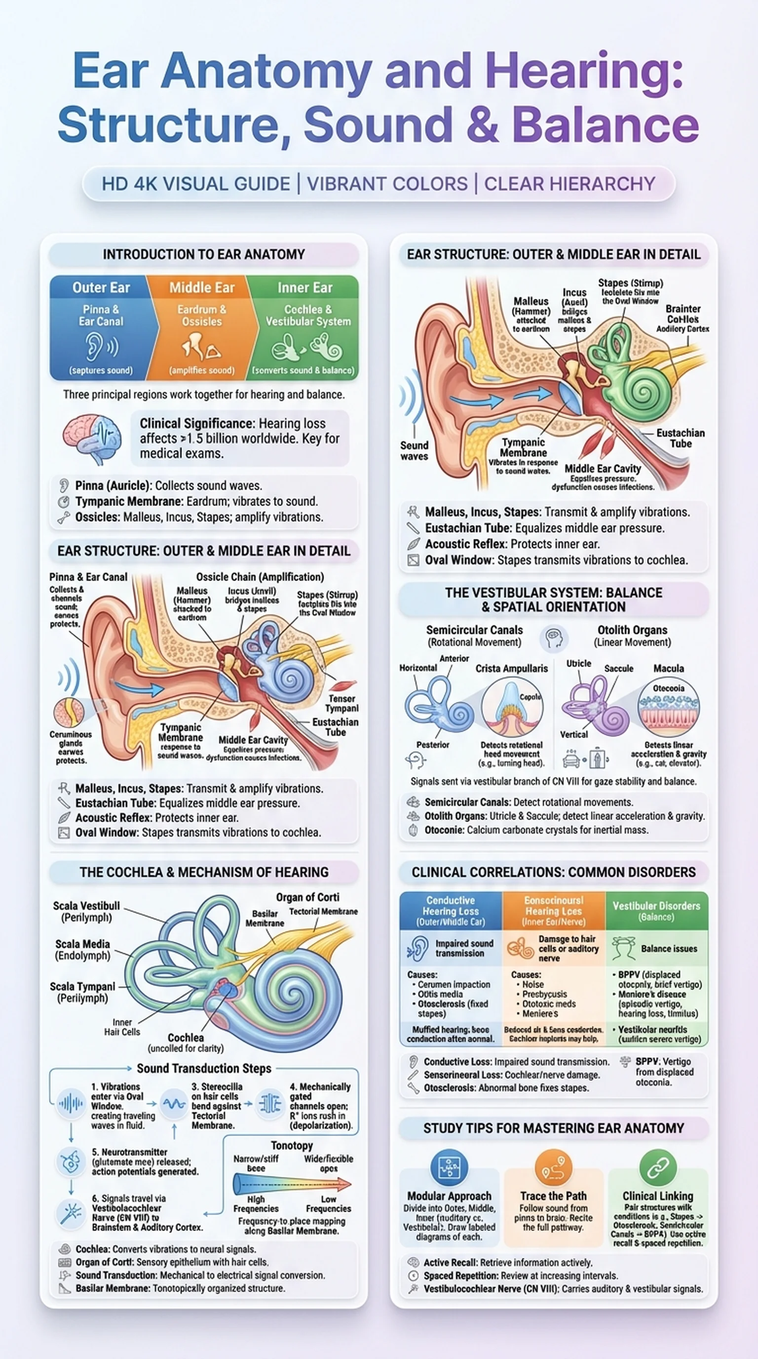
Introduction to Ear Anatomy

Ear anatomy is a rich and multifaceted subject that encompasses not only the structures responsible for hearing but also those that govern balance and spatial orientation. The human ear is divided into three principal regions: the outer ear, the middle ear, and the inner ear. Each region contains specialized structures that work in concert to capture sound waves from the environment, convert them into mechanical vibrations, transform those vibrations into electrical signals, and relay those signals to the auditory cortex of the brain.

Understanding ear structure is essential for students of anatomy, audiology, otolaryngology, and neuroscience. The ear appears on virtually every major medical and health sciences examination, from the USMLE and MCAT to audiology certification boards. Its clinical significance is vast: hearing loss affects over 1.5 billion people worldwide, and vestibular disorders are among the most common complaints seen in primary care settings.

The outer ear consists of the pinna (auricle) and the external auditory canal, which together funnel sound toward the tympanic membrane. The middle ear is an air-filled cavity containing the three smallest bones in the human body, the ossicles, which mechanically amplify sound vibrations. The inner ear houses the cochlea, the spiral-shaped organ responsible for sound transduction, and the vestibular system, which detects head position and movement. This introductory overview of ear anatomy sets the stage for a deeper exploration of how each component contributes to the remarkable processes of hearing and balance.

Key Terms

Ear Anatomy

The study of the structural components of the ear, divided into the outer ear, middle ear, and inner ear, each serving distinct roles in hearing and balance.

Pinna (Auricle)

The external, visible portion of the ear that collects and directs sound waves into the external auditory canal.

Tympanic Membrane

The eardrum; a thin, semitransparent membrane separating the outer ear from the middle ear that vibrates in response to sound waves.

Ossicles

The three small bones of the middle ear (malleus, incus, stapes) that transmit and amplify vibrations from the tympanic membrane to the inner ear.

Ear Structure: Outer and Middle Ear in Detail

A comprehensive grasp of ear structure begins with the outer ear. The pinna is composed of elastic cartilage covered by skin, and its complex folds and ridges (including the helix, antihelix, tragus, and concha) are shaped to collect sound waves and channel them efficiently into the external auditory canal. The canal itself is approximately 2.5 centimeters long in adults and is lined with ceruminous glands that produce earwax (cerumen) to protect against debris and infection. Sound waves travel through the canal and strike the tympanic membrane, causing it to vibrate.

The middle ear, also called the tympanic cavity, is an air-filled space within the temporal bone. Its medial wall borders the inner ear, while its lateral wall is formed by the tympanic membrane. The three ossicles span this cavity in a chain: the malleus is attached to the inner surface of the tympanic membrane, the incus bridges the malleus and the stapes, and the footplate of the stapes fits into the oval window of the inner ear. This ossicular chain amplifies sound pressure by approximately 20-fold through two mechanisms: the lever action of the ossicles and the area ratio between the large tympanic membrane and the small oval window.

Two muscles within the middle ear protect against dangerously loud sounds. The tensor tympani, innervated by the trigeminal nerve, tenses the tympanic membrane, while the stapedius, innervated by the facial nerve, stabilizes the stapes. Together they constitute the acoustic reflex. The middle ear also communicates with the nasopharynx via the Eustachian tube (pharyngotympanic tube), which equalizes pressure across the tympanic membrane. Dysfunction of the Eustachian tube is a common cause of middle ear infections (otitis media), particularly in children, making knowledge of this ear structure clinically critical.

Key Terms

Ear Structure

The physical arrangement of the outer, middle, and inner ear, including bones, membranes, and fluid-filled chambers that enable hearing and balance.

Malleus, Incus, Stapes

The three ossicles of the middle ear; the malleus (hammer), incus (anvil), and stapes (stirrup) form a chain that transmits vibrations to the inner ear.

Eustachian Tube

A canal connecting the middle ear to the nasopharynx that equalizes air pressure on both sides of the tympanic membrane.

Acoustic Reflex

The involuntary contraction of the stapedius and tensor tympani muscles in response to loud sounds, protecting the inner ear from damage.

Oval Window

A membrane-covered opening in the medial wall of the middle ear where the stapes footplate transmits vibrations into the fluid-filled cochlea.

The Cochlea and the Mechanism of Hearing

The cochlea is the snail-shaped, fluid-filled organ of the inner ear that is responsible for converting mechanical vibrations into the electrical signals we perceive as hearing. Coiled approximately two and a half turns around a bony core called the modiolus, the cochlea is divided into three parallel chambers by two membranes. The scala vestibuli (upper chamber) and scala tympani (lower chamber) are filled with perilymph, a fluid similar in composition to extracellular fluid. Between them lies the scala media (cochlear duct), filled with endolymph, a potassium-rich fluid essential for the process of sound transduction.

The organ of Corti, situated on the basilar membrane within the scala media, is the sensory epithelium of hearing. It contains approximately 15,000 hair cells arranged in one row of inner hair cells and three rows of outer hair cells. When sound-induced vibrations enter the cochlea through the oval window, they create traveling waves along the basilar membrane. The basilar membrane is tonotopically organized: the base is narrow and stiff, responding to high-frequency sounds, while the apex is wide and flexible, responding to low-frequency sounds. This frequency-to-place mapping is one of the most important principles in auditory physiology.

Sound transduction occurs when displacement of the basilar membrane causes the stereocilia atop the hair cells to bend against the tectorial membrane. Mechanically gated ion channels at the tips of the stereocilia open, allowing potassium ions from the endolymph to rush into the hair cell, depolarizing it. This depolarization triggers the release of glutamate at the synapse between the inner hair cell and afferent fibers of the cochlear branch of cranial nerve VIII (the vestibulocochlear nerve). The resulting action potentials travel to the brainstem, and the brain interprets these signals as sound. This elegant mechanism of hearing allows humans to detect frequencies ranging from approximately 20 Hz to 20,000 Hz.

Key Terms

Cochlea

The spiral-shaped, fluid-filled organ of the inner ear containing the organ of Corti, responsible for converting mechanical vibrations into neural signals for hearing.

Organ of Corti

The sensory epithelium within the cochlea containing inner and outer hair cells that transduce sound vibrations into electrical signals.

Sound Transduction

The process by which mechanical sound vibrations are converted into electrical nerve impulses in the cochlear hair cells.

Basilar Membrane

The membrane within the cochlea upon which the organ of Corti rests; its tonotopic organization maps sound frequency to position along its length.

Tonotopy

The spatial arrangement of sound frequency processing along the basilar membrane, with high frequencies at the base and low frequencies at the apex.

The Vestibular System: Balance and Spatial Orientation

The vestibular system is the sensory apparatus of the inner ear responsible for detecting head position, linear acceleration, and rotational movement. While the cochlea handles hearing, the vestibular system ensures that we can maintain balance, stabilize our gaze during head movements, and navigate through three-dimensional space. The vestibular system comprises five sensory organs: three semicircular canals and two otolith organs (the utricle and saccule).

The three semicircular canals are oriented roughly perpendicular to one another, corresponding to the three planes of space: horizontal (lateral), anterior (superior), and posterior. Each canal is filled with endolymph and contains a dilated region called the ampulla. Within the ampulla sits the crista ampullaris, a ridge of hair cells embedded in a gelatinous structure called the cupula. When the head rotates, inertia causes the endolymph to lag behind, deflecting the cupula and bending the stereocilia of the hair cells. This deflection either depolarizes or hyperpolarizes the hair cells depending on the direction of movement, signaling rotational acceleration to the brain via the vestibular branch of cranial nerve VIII.

The otolith organs detect linear acceleration and static head tilt relative to gravity. The utricle is oriented horizontally and senses movements such as riding in a car, while the saccule is oriented vertically and senses movements like riding in an elevator. Both contain a sensory epithelium called the macula, where hair cells are embedded in a gelatinous layer topped with calcium carbonate crystals called otoconia (otoliths). When the head tilts or accelerates linearly, the heavy otoconia shift, bending the hair cell stereocilia and generating signals transmitted along the vestibular nerve. Dysfunction of the vestibular system leads to vertigo, nystagmus, and balance disorders, underscoring its critical importance in daily life and clinical medicine.

Key Terms

Vestibular System

The sensory system within the inner ear that detects head position and movement, comprising three semicircular canals and two otolith organs (utricle and saccule).

Semicircular Canals

Three fluid-filled loops in the inner ear oriented in different planes that detect rotational head movements.

Otolith Organs

The utricle and saccule; vestibular structures containing calcium carbonate crystals that detect linear acceleration and gravitational orientation.

Crista Ampullaris

The sensory receptor within the ampulla of each semicircular canal, containing hair cells and a gelatinous cupula that responds to rotational movement.

Otoconia

Tiny calcium carbonate crystals on the maculae of the utricle and saccule that add inertial mass, enabling detection of linear acceleration and head tilt.

Clinical Correlations: Common Disorders of the Ear

A solid understanding of ear anatomy translates directly into clinical competence, as disorders of the ear are among the most frequently encountered conditions in medicine. Conductive hearing loss results from impaired sound transmission through the outer or middle ear. Common causes include cerumen impaction, tympanic membrane perforation, otitis media with effusion, and otosclerosis, a condition in which abnormal bone growth fixes the stapes footplate to the oval window. Patients with conductive hearing loss often report muffled hearing but typically retain a normal ability to hear bone-conducted sound, a distinction exploited by the Rinne and Weber tuning fork tests.

Sensorineural hearing loss arises from damage to the cochlea or the auditory nerve. Noise-induced hearing loss, presbycusis (age-related hearing loss), ototoxic medications (such as aminoglycosides and cisplatin), and Meniere's disease are common causes. In sensorineural hearing loss, both air and bone conduction are reduced because the problem lies in sound transduction or neural transmission, not mechanical conduction. Cochlear implants, which bypass damaged hair cells and directly stimulate the cochlear nerve, have revolutionized treatment for severe sensorineural hearing loss.

Vestibular disorders represent another major category of ear pathology. Benign paroxysmal positional vertigo (BPPV) occurs when displaced otoconia enter a semicircular canal, causing brief episodes of intense vertigo with head position changes. Meniere's disease involves endolymphatic hydrops, leading to episodic vertigo, fluctuating hearing loss, tinnitus, and aural fullness. Vestibular neuritis, an inflammation of the vestibular nerve, causes sudden, severe vertigo lasting days. Each of these conditions maps to a specific anatomical structure within the ear, reinforcing why detailed knowledge of ear anatomy is indispensable for clinicians evaluating hearing and balance complaints.

Key Terms

Conductive Hearing Loss

Hearing loss caused by impaired transmission of sound through the outer or middle ear structures.

Sensorineural Hearing Loss

Hearing loss resulting from damage to the cochlea (hair cells) or the auditory nerve, impairing sound transduction or neural transmission.

Otosclerosis

A condition of abnormal bone remodeling in the middle ear that fixes the stapes footplate, causing progressive conductive hearing loss.

Benign Paroxysmal Positional Vertigo (BPPV)

A vestibular disorder caused by displaced otoconia in the semicircular canals, producing brief episodes of vertigo triggered by head position changes.

Study Tips for Mastering Ear Anatomy and Hearing

Ear anatomy and hearing physiology can seem overwhelming because of the intricate structures and the dual functions of audition and balance. However, a systematic study approach transforms this complexity into manageable, interconnected concepts. Here are proven strategies for mastering ear structure and the mechanisms of hearing.

First, divide your study into three clear modules: outer ear, middle ear, and inner ear. Within the inner ear, further separate the auditory (cochlear) and vestibular systems. This compartmentalization prevents confusion and allows you to focus on one functional unit at a time. Create labeled diagrams for each module. Drawing the cochlea in cross-section with the scala vestibuli, scala media, and scala tympani clearly marked is particularly high-yield for exams.

Second, trace the path of sound from source to perception. Start with sound waves entering the pinna, traveling through the external auditory canal, vibrating the tympanic membrane, being amplified by the ossicles, entering the cochlea through the oval window, creating traveling waves on the basilar membrane, triggering sound transduction in the organ of Corti, and generating action potentials that travel via the vestibulocochlear nerve to the auditory cortex. Being able to recite this complete pathway demonstrates mastery of both ear anatomy and auditory physiology.

Third, pair each structure with its associated clinical condition. The stapes maps to otosclerosis. The cochlear hair cells map to noise-induced hearing loss. The semicircular canals map to BPPV. This clinical-anatomical linking strategy is exactly how exam questions are designed. Finally, leverage active learning tools like LectureScribe to create spaced repetition flashcards and slide presentations from your lecture notes. Consistent, spaced review of ear anatomy and hearing concepts builds durable long-term memory that holds up under exam pressure.

Key Terms

Active Recall

A study technique where learners actively retrieve information from memory rather than passively reviewing notes.

Spaced Repetition

A learning strategy that involves reviewing material at increasing intervals to strengthen long-term memory retention.

Vestibulocochlear Nerve

Cranial nerve VIII, which carries both auditory signals from the cochlea and vestibular signals from the semicircular canals and otolith organs to the brainstem.

Frequently Asked Questions

What are the main parts of ear anatomy?

Ear anatomy is divided into three regions: the outer ear (pinna and external auditory canal), the middle ear (tympanic membrane, ossicles, and Eustachian tube), and the inner ear (cochlea for hearing and the vestibular system for balance). Each region plays a specific role in capturing, transmitting, and processing sound.

How does the ear structure enable hearing?

The ear structure enables hearing through a chain of events: the pinna collects sound waves, the tympanic membrane converts them to vibrations, the ossicles amplify these vibrations, and the cochlea converts them into electrical signals through sound transduction in the organ of Corti. These signals travel via the vestibulocochlear nerve to the auditory cortex.

What is the cochlea and what does it do?

The cochlea is a snail-shaped, fluid-filled organ in the inner ear that converts mechanical sound vibrations into electrical nerve signals. It contains the organ of Corti with hair cells that perform sound transduction. The cochlea's basilar membrane is tonotopically organized, mapping different sound frequencies to different locations.

What is the vestibular system?

The vestibular system is the balance apparatus of the inner ear, consisting of three semicircular canals that detect rotational movement and two otolith organs (utricle and saccule) that detect linear acceleration and head tilt relative to gravity. It sends signals via the vestibular nerve to the brainstem for balance control.

How does sound transduction work in the ear?

Sound transduction occurs in the cochlea when vibrations create traveling waves along the basilar membrane. These waves bend the stereocilia of hair cells against the tectorial membrane, opening mechanically gated ion channels. Potassium influx depolarizes the hair cell, triggering glutamate release and generating action potentials in the cochlear nerve fibers.

What is the difference between conductive and sensorineural hearing loss?

Conductive hearing loss results from impaired sound transmission through the outer or middle ear, such as from otosclerosis or tympanic membrane perforation. Sensorineural hearing loss results from damage to the cochlea or auditory nerve, as in noise-induced hearing loss. Conductive loss affects air conduction only, while sensorineural loss affects both air and bone conduction.

How do the semicircular canals detect movement?

The three semicircular canals are oriented in perpendicular planes and filled with endolymph. When the head rotates, the endolymph lags due to inertia, deflecting the cupula in the ampulla and bending hair cell stereocilia. This generates electrical signals sent via the vestibular nerve to the brainstem, encoding the direction and speed of head rotation.

Study Anatomy Smarter

Upload your lecture notes and get AI-generated flashcards, quizzes, infographics, and study guides tailored to your curriculum.

Try LectureScribe Free

Create Study Materials

Transform your Anatomy lectures into flashcards, quizzes, and visual study guides with AI.

  • AI-generated flashcards & quizzes
  • Visual infographics from notes
  • Interactive study shorts
Start Studying Free

Quick Facts

SubjectAnatomy
Word Count2,500+
Slides5
InfographicIncluded
AuthorLectureScribe

Master Anatomy with LectureScribe

Upload your lecture notes and get AI-powered flashcards, quizzes, infographics, and study guides in minutes.