LectureScribe Logo
lecturescribe.io

LOCK IN VALENTINE PRICING BEFORE SALE ENDS!

Use code — New joiners get 40% OFF on annual plans

Plans will have higher prices after this sale ends

0days
:
0hours
:
0minutes
:
0seconds
Biology2,500+ words5 slides
📦

Golgi Apparatus Function: Structure, Protein Processing, and Cell Biology

Explore the Golgi apparatus function in cell biology. Learn how the Golgi body processes, sorts, and ships proteins, and discover disorders linked to the Golgi complex.

L
LectureScribeAI-Powered Study Platform

Study Infographic

Diagram of Golgi apparatus structure and function, illustrating the flow of proteins from the ER through cis, medial, and trans cisternae to their final destinations.

Download
Golgi apparatus function diagram showing cis, medial, and trans compartments with protein processing pathway

Interactive Study Short

Swipe through 5 slides about Golgi Apparatus Function: Structure, Protein Processing, and Cell Biology

1 / 5

What Is the Golgi Apparatus?

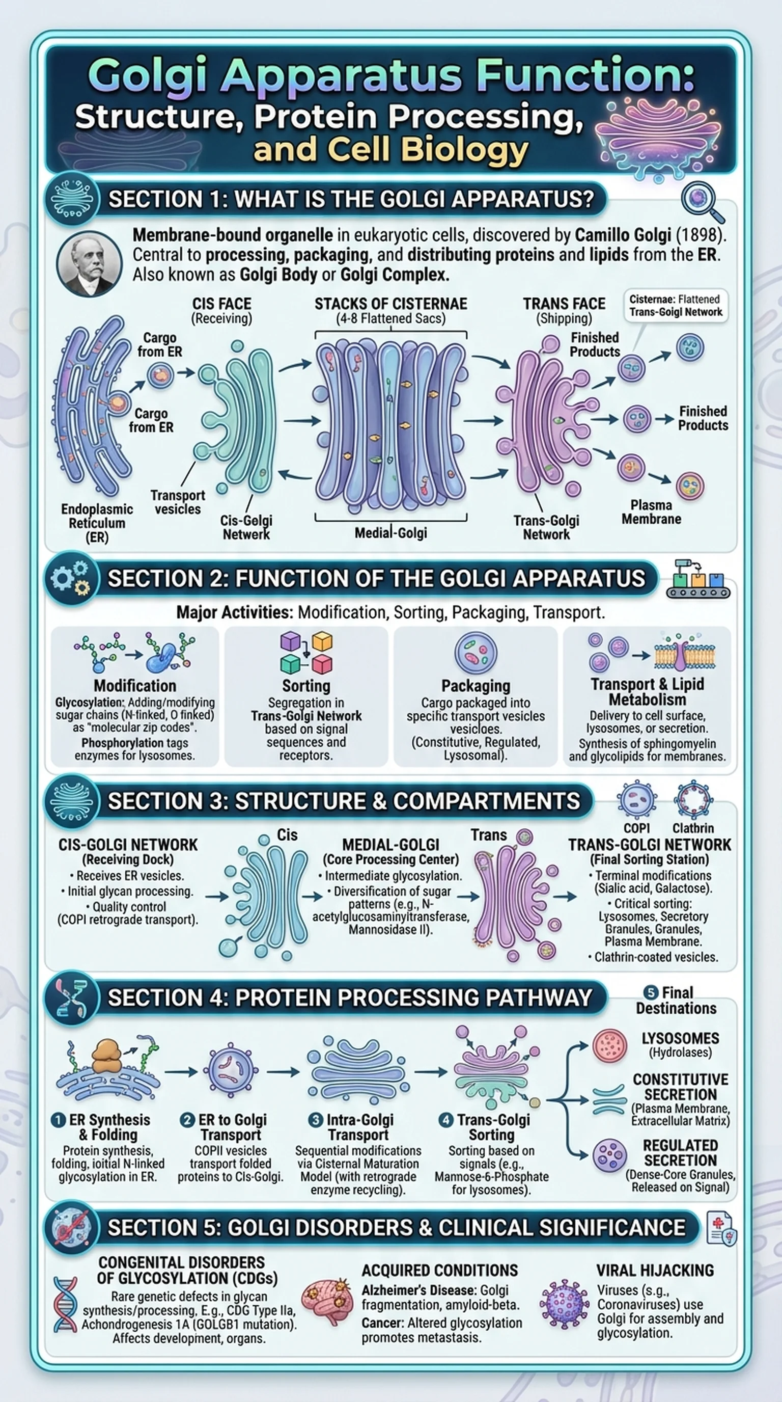
The Golgi apparatus is a membrane-bound organelle found in virtually all eukaryotic cells. First described by the Italian physician Camillo Golgi in 1898, the Golgi apparatus plays a central role in processing, packaging, and distributing proteins and lipids produced by the endoplasmic reticulum. It is sometimes referred to as the Golgi body or the Golgi complex, and all three names describe the same organelle with the same essential functions.

Structurally, the Golgi apparatus consists of a series of flattened, stacked membrane sacs called cisternae. A typical mammalian cell contains between 40 and 100 cisternae organized into four to eight stacks. Each stack has a distinct polarity: the cis face (receiving side) is oriented toward the endoplasmic reticulum, while the trans face (shipping side) faces the plasma membrane. Transport vesicles bud from the ER and fuse with the cis-Golgi, delivering their cargo for processing. After modification, finished products exit from the trans-Golgi network in vesicles destined for the cell surface, lysosomes, or secretory pathways.

The Golgi apparatus is especially prominent in cells that specialize in secretion, such as goblet cells in the intestinal lining and plasma cells in the immune system. In plant cells, the Golgi body also synthesizes polysaccharides for the cell wall. Understanding the Golgi apparatus is fundamental for students of cell biology, biochemistry, and medicine, as its dysfunction is implicated in numerous diseases.

Key Terms

Golgi Apparatus

A membrane-bound organelle composed of stacked cisternae that modifies, sorts, and packages proteins and lipids for transport within or outside the cell.

Golgi Body

An alternative name for the Golgi apparatus, commonly used in introductory biology courses.

Golgi Complex

Another name for the Golgi apparatus, emphasizing its structural complexity of multiple cisternae and associated vesicles.

Cisternae

Flattened, membrane-bound sacs that make up the stacked structure of the Golgi apparatus.

Trans-Golgi Network

The sorting and shipping station at the trans face of the Golgi apparatus, where modified proteins are packaged into vesicles for their final destinations.

Function of the Golgi Apparatus in the Cell

The Golgi apparatus function can be understood through four major activities: modification, sorting, packaging, and transport of macromolecules. Each of these activities is essential for maintaining cellular organization and enabling communication between cells.

Protein modification is perhaps the best-known Golgi apparatus function. As proteins move through the cisternae from the cis to the trans face, resident enzymes add, remove, or modify sugar chains in a process called glycosylation. N-linked oligosaccharides that were initially added in the endoplasmic reticulum are trimmed and remodeled, and O-linked sugars may be attached to serine or threonine residues. These glycosylation patterns serve as molecular zip codes, directing proteins to their correct destinations. Phosphorylation of mannose residues, for example, tags lysosomal enzymes for delivery to lysosomes. Without proper Golgi apparatus function, these enzymes would be secreted outside the cell instead.

Sorting and packaging occur primarily in the trans-Golgi network. Here, modified proteins and lipids are segregated into different transport vesicles based on signal sequences and receptor interactions. Constitutive secretory vesicles carry cargo that is continuously released from the cell, while regulated secretory vesicles store their contents until an extracellular signal triggers exocytosis. The Golgi body also produces lysosomes by packaging hydrolytic enzymes into vesicles coated with specific membrane markers.

Lipid metabolism is another critical function of the Golgi complex. Sphingomyelin and glycolipids are synthesized in the Golgi from ceramide precursors delivered from the ER. These lipids are incorporated into the plasma membrane, where they play roles in cell signaling and membrane structure. The Golgi apparatus function in lipid processing is therefore integral to maintaining the composition and asymmetry of cellular membranes.

Key Terms

Glycosylation

The enzymatic process of adding sugar chains to proteins or lipids, a primary modification carried out in the Golgi apparatus.

Constitutive Secretion

The continuous, unregulated release of vesicle contents from the cell, mediated by the Golgi apparatus.

Regulated Secretion

The signal-dependent release of stored vesicle contents, requiring an external trigger such as a hormone or neurotransmitter.

Exocytosis

The process by which vesicles fuse with the plasma membrane to release their contents outside the cell.

Sphingomyelin

A phospholipid synthesized in the Golgi apparatus that is a major component of the plasma membrane and myelin sheaths.

Structure of the Golgi Apparatus: Cis, Medial, and Trans Compartments

The structure of the Golgi apparatus is intimately linked to its function. The organelle is organized into three functionally distinct regions: the cis-Golgi (nearest the ER), the medial-Golgi (middle cisternae), and the trans-Golgi (nearest the plasma membrane). Each compartment contains a unique set of enzymes that carry out specific modifications in an assembly-line fashion as cargo moves from cis to trans.

The cis-Golgi network, also known as the cis-Golgi reticulum, serves as the receiving dock. Transport vesicles budding from the ER fuse with this compartment, delivering newly synthesized proteins and lipids. The cis-Golgi is responsible for the initial steps of glycan processing, including the removal of specific mannose residues from N-linked oligosaccharides. It also performs quality control, sending misfolded proteins back to the ER through retrograde transport vesicles coated with COPI proteins.

The medial-Golgi cisternae contain enzymes such as N-acetylglucosaminyltransferase and mannosidase II, which further trim and elaborate the carbohydrate chains on glycoproteins. This is where the Golgi complex begins to diversify the glycosylation patterns that will ultimately determine each protein's destination and function. The medial compartment is often considered the core processing center of the Golgi body.

The trans-Golgi and trans-Golgi network represent the final processing and sorting stations. Terminal sugar modifications, including the addition of sialic acid and galactose, occur here. The trans-Golgi network is where the critical sorting decisions are made: proteins are directed toward the plasma membrane, endosomes, lysosomes, or secretory granules. Clathrin-coated vesicles bud from this region to carry lysosomal enzymes, while other vesicle types handle constitutive and regulated secretory pathways. The structural polarity of the Golgi apparatus thus reflects a directional processing pipeline essential for proper cell function.

Key Terms

Cis-Golgi Network

The receiving compartment of the Golgi apparatus closest to the ER, where initial glycan processing and quality control occur.

Medial-Golgi

The middle cisternae of the Golgi apparatus where intermediate glycosylation modifications take place.

COPI Vesicles

Coat protein complex I vesicles that mediate retrograde transport from the Golgi back to the ER.

Clathrin-Coated Vesicles

Transport vesicles surrounded by a clathrin protein lattice, involved in trafficking from the trans-Golgi network to endosomes and lysosomes.

The Golgi Apparatus and Protein Processing Pathway

The journey of a protein through the Golgi apparatus is a carefully orchestrated process that begins at the ribosome and ends with the protein arriving at its functional destination. Understanding this pathway is essential for appreciating the full scope of Golgi apparatus function in the secretory system.

Protein synthesis begins on ribosomes attached to the rough endoplasmic reticulum. As the polypeptide chain grows, it is threaded into the ER lumen, where chaperone proteins assist with folding and initial quality control. Core N-linked glycosylation occurs co-translationally in the ER, adding a 14-sugar oligosaccharide to asparagine residues. Once properly folded, proteins are packaged into COPII-coated transport vesicles that bud from specialized ER exit sites and travel to the cis face of the Golgi body.

Within the Golgi complex, proteins undergo sequential modifications as they traverse from cis to trans cisternae. The two prevailing models for intra-Golgi transport are the vesicular transport model, in which cargo moves via shuttling vesicles between stable cisternae, and the cisternal maturation model, in which entire cisternae progressively mature from cis to trans while resident Golgi enzymes are recycled backward via retrograde vesicles. Current evidence supports a modified version of cisternal maturation with some vesicular transport components.

As proteins reach the trans-Golgi network, they receive final modifications and are sorted based on signal sequences. Mannose-6-phosphate tags direct hydrolases to lysosomes. Secretory proteins lacking specific retention signals follow the default constitutive pathway to the cell surface. Regulated secretory proteins aggregate in the trans-Golgi network and are stored in dense-core granules until the appropriate stimulus triggers their release. This elaborate sorting machinery ensures that each protein produced by the cell reaches the correct compartment, and it illustrates why the Golgi apparatus function is critical for cellular homeostasis.

Key Terms

COPII Vesicles

Coat protein complex II vesicles that mediate anterograde transport from the ER to the Golgi apparatus.

Cisternal Maturation Model

A model of Golgi transport in which cisternae progressively mature from cis to trans, with Golgi enzymes recycled backward via retrograde vesicles.

Mannose-6-Phosphate

A molecular tag added to lysosomal enzymes in the Golgi apparatus, directing them to lysosomes via receptor-mediated sorting.

Signal Sequence

A short peptide sequence on a protein that directs it to a specific cellular compartment, recognized by sorting machinery in the Golgi.

Dense-Core Granules

Membrane-bound storage vesicles in the regulated secretory pathway that release their contents upon extracellular stimulation.

Golgi Apparatus Disorders and Clinical Significance

Disruption of Golgi apparatus function leads to a class of diseases collectively known as congenital disorders of glycosylation (CDGs). These rare genetic conditions result from mutations in enzymes responsible for glycan synthesis or processing within the Golgi complex and endoplasmic reticulum. Patients with CDGs may present with developmental delays, neurological abnormalities, liver dysfunction, and immune deficiencies, illustrating the far-reaching consequences of impaired Golgi body function.

One well-characterized example is CDG type IIa, caused by a deficiency in N-acetylglucosaminyltransferase II, an enzyme localized to the medial-Golgi. Without this enzyme, glycoproteins lack proper complex-type glycans, leading to multi-system organ dysfunction. Another example is the hereditary disorder known as achondrogenesis type 1A, linked to mutations in the GOLGB1 gene that encodes a Golgi matrix protein essential for maintaining cisterna structure. When the architecture of the Golgi apparatus is compromised, cartilage and bone development are severely affected.

Beyond genetic diseases, the Golgi apparatus plays important roles in acquired conditions. In Alzheimer's disease, fragmentation of the Golgi complex has been observed in neurons, potentially contributing to impaired protein trafficking and the accumulation of amyloid-beta plaques. In cancer biology, alterations in Golgi-mediated glycosylation can promote tumor invasion and metastasis by changing the surface glycoprotein profiles of malignant cells. Researchers are actively investigating whether targeting Golgi apparatus function could yield new therapeutic strategies for these conditions.

Viral infections also exploit the Golgi body. Many enveloped viruses, including coronaviruses and flaviviruses, hijack the Golgi complex to assemble and glycosylate their surface proteins before budding from the host cell. Understanding how viruses co-opt Golgi apparatus function is crucial for developing antiviral drugs that interrupt viral assembly pathways. These clinical connections underscore the importance of the Golgi apparatus not only in basic cell biology but also in medicine and pharmacology.

Key Terms

Congenital Disorders of Glycosylation (CDGs)

A group of inherited metabolic disorders caused by defects in glycan synthesis or processing, often involving Golgi apparatus enzymes.

GOLGB1

A gene encoding a Golgi matrix protein (giantin) essential for maintaining the structural integrity of the Golgi apparatus.

Golgi Fragmentation

The pathological breakdown of the Golgi apparatus into smaller vesicles, observed in neurodegenerative diseases and some cancers.

Amyloid-Beta Plaques

Abnormal protein aggregates found in the brains of Alzheimer's disease patients, whose formation may be linked to impaired Golgi trafficking.

Frequently Asked Questions

What is the main function of the Golgi apparatus?

The main Golgi apparatus function is to modify, sort, and package proteins and lipids received from the endoplasmic reticulum for transport to their final destinations within or outside the cell. This includes glycosylation, phosphorylation, and vesicle-mediated trafficking.

What is the difference between the Golgi body, Golgi complex, and Golgi apparatus?

There is no difference. Golgi body, Golgi complex, and Golgi apparatus are three names for the same organelle. All refer to the stacked membrane cisternae that process and distribute proteins and lipids in eukaryotic cells.

Where is the Golgi apparatus located in the cell?

The Golgi apparatus is typically located near the nucleus and the centrosome in animal cells. Its cis face is oriented toward the endoplasmic reticulum, and its trans face is directed toward the plasma membrane, reflecting the directional flow of protein processing.

What happens when the Golgi apparatus malfunctions?

When the Golgi apparatus malfunctions, proteins and lipids cannot be properly modified or sorted. This can lead to congenital disorders of glycosylation, impaired secretion, and has been linked to neurodegenerative diseases such as Alzheimer's disease and to cancer progression.

How does the Golgi apparatus function in protein glycosylation?

The Golgi apparatus carries out sequential glycosylation as proteins move from cis to trans cisternae. Enzymes in each compartment trim, add, and modify sugar chains on glycoproteins. These glycan modifications serve as signals that direct proteins to their correct cellular destinations.

Why is the Golgi body important for secretory cells?

The Golgi body is especially important for secretory cells because it packages large quantities of proteins into secretory vesicles for export. Cells like goblet cells and plasma cells rely heavily on the Golgi complex to process and release mucus and antibodies, respectively.

How do viruses exploit the Golgi complex?

Enveloped viruses such as coronaviruses hijack the Golgi complex to glycosylate and assemble their surface proteins. By co-opting the Golgi apparatus function, viruses ensure their envelope proteins are properly modified before the viral particles bud from the host cell.

Study Biology Smarter

Upload your lecture notes and get AI-generated flashcards, quizzes, infographics, and study guides tailored to your curriculum.

Try LectureScribe Free

Create Study Materials

Transform your Biology lectures into flashcards, quizzes, and visual study guides with AI.

  • AI-generated flashcards & quizzes
  • Visual infographics from notes
  • Interactive study shorts
Start Studying Free

Quick Facts

SubjectBiology
Word Count2,500+
Slides5
InfographicIncluded
AuthorLectureScribe

Master Biology with LectureScribe

Upload your lecture notes and get AI-powered flashcards, quizzes, infographics, and study guides in minutes.