LectureScribe Logo
lecturescribe.io

LOCK IN VALENTINE PRICING BEFORE SALE ENDS!

Use code — New joiners get 40% OFF on annual plans

Plans will have higher prices after this sale ends

0days
:
0hours
:
0minutes
:
0seconds
Biochemistry2,500+ words5 slides
🩸

Hemoglobin and Oxygen Binding: Cooperative Binding & Bohr Effect

Understand hemoglobin and oxygen binding, cooperative binding, the Bohr effect, and the oxygen dissociation curve. A complete biochemistry guide for students preparing for the MCAT and USMLE.

L
LectureScribeAI-Powered Study Platform

Study Infographic

Hemoglobin oxygen dissociation curve illustrating cooperative binding, allosteric shifts caused by pH and CO2 (Bohr effect), and comparison with myoglobin's hyperbolic curve.

Download
Hemoglobin oxygen dissociation curve showing sigmoidal shape, T and R state transitions, and rightward shift caused by the Bohr effect

Interactive Study Short

Swipe through 5 slides about Hemoglobin and Oxygen Binding: Cooperative Binding & Bohr Effect

1 / 5

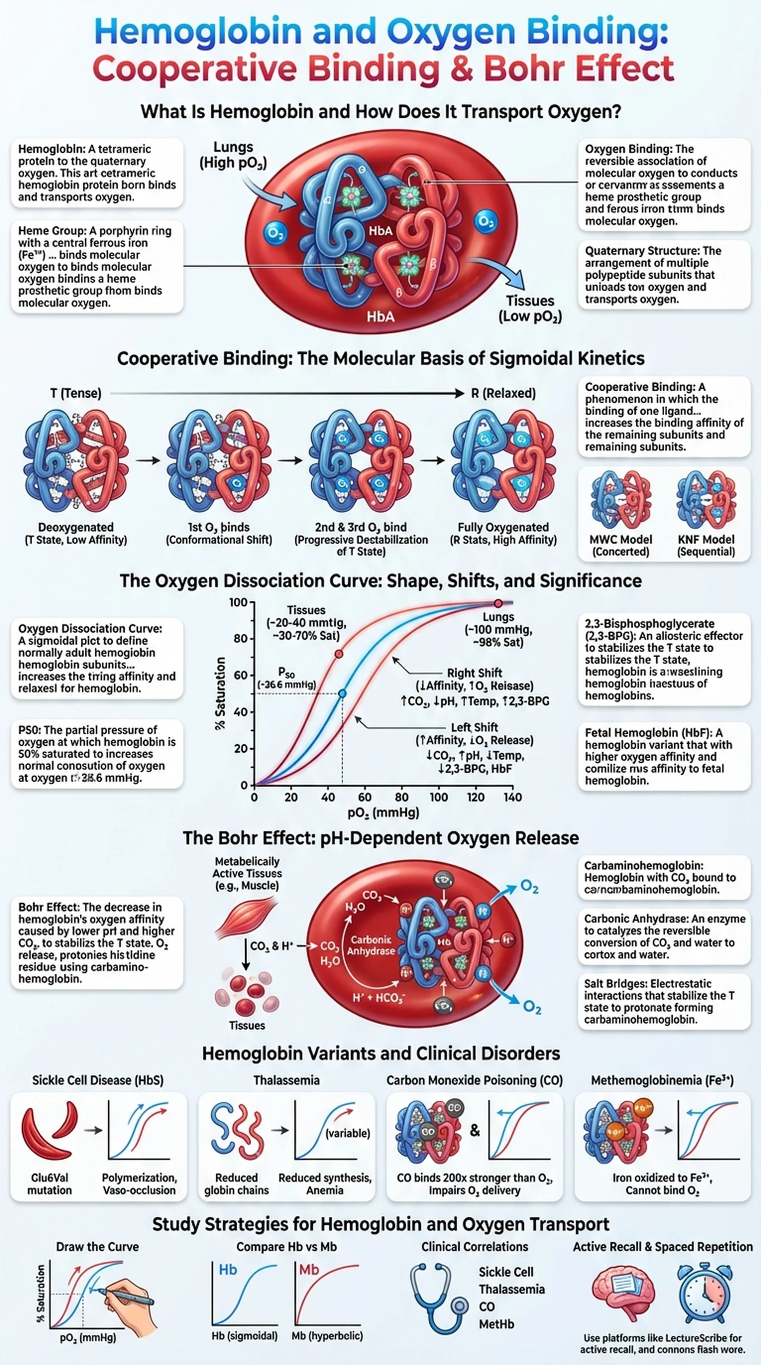
What Is Hemoglobin and How Does It Transport Oxygen?

Hemoglobin is a tetrameric metalloprotein found in red blood cells (erythrocytes) that is responsible for transporting oxygen from the lungs to the tissues and facilitating the return of carbon dioxide to the lungs for exhalation. Each hemoglobin molecule consists of four polypeptide subunits, two alpha chains and two beta chains in adult hemoglobin (HbA), and each subunit contains a heme prosthetic group with a central iron atom in the ferrous (Fe2+) state. It is this iron atom that reversibly binds one molecule of O2, giving each hemoglobin tetramer the capacity to carry up to four oxygen molecules simultaneously.

The ability of hemoglobin to bind and release oxygen efficiently is essential for aerobic life. In the lungs, where the partial pressure of oxygen is high, hemoglobin becomes nearly fully saturated with O2. In the peripheral tissues, where oxygen tension is lower and metabolic demand is high, hemoglobin releases its oxygen cargo to supply the cells. This loading and unloading behavior is not a simple on-off switch; instead, it is governed by a sophisticated set of molecular interactions that include cooperative binding, allosteric regulation, and sensitivity to local chemical conditions such as pH, CO2 concentration, and temperature.

Understanding oxygen binding by hemoglobin is foundational for students of biochemistry, physiology, and medicine. Abnormalities in hemoglobin structure or function underlie diseases such as sickle cell anemia, thalassemia, and carbon monoxide poisoning. The principles governing hemoglobin's behavior also illustrate broader biochemical concepts including allostery, protein quaternary structure, and the relationship between structure and function that are central to molecular biology.

Key Terms

Hemoglobin

A tetrameric protein in red blood cells composed of four subunits, each containing a heme group, that reversibly binds and transports oxygen.

Heme Group

A porphyrin ring with a central ferrous iron (Fe2+) atom that directly binds molecular oxygen in each hemoglobin subunit.

Oxygen Binding

The reversible association of molecular oxygen with the iron atom in hemoglobin's heme group, enabling oxygen transport from lungs to tissues.

Quaternary Structure

The arrangement of multiple polypeptide subunits into a functional protein complex, as seen in hemoglobin's alpha2-beta2 tetramer.

Cooperative Binding: The Molecular Basis of Sigmoidal Kinetics

Cooperative binding is the hallmark feature that distinguishes hemoglobin from simpler oxygen-carrying proteins like myoglobin. In cooperative binding, the attachment of the first oxygen molecule to one subunit of hemoglobin increases the affinity of the remaining subunits for subsequent oxygen molecules. Conversely, the release of the first oxygen molecule facilitates the release of the others. This positive cooperativity means that hemoglobin transitions between two conformational states: the tense (T) state, which has low oxygen affinity, and the relaxed (R) state, which has high oxygen affinity.

When hemoglobin is fully deoxygenated, all four subunits are in the T state. The binding of the first O2 molecule to one subunit triggers a conformational shift in the iron atom, pulling it into the plane of the porphyrin ring and repositioning the proximal histidine residue. This local structural change is transmitted through subunit interfaces to the other subunits, progressively destabilizing the T state and favoring the transition to the R state. By the time the third or fourth oxygen molecule binds, the protein is predominantly in the R state and oxygen affinity is at its maximum.

The Monod-Wyman-Changeux (MWC) concerted model and the Koshland-Nemethy-Filmer (KNF) sequential model are the two classical frameworks used to describe cooperative binding. The MWC model proposes that all subunits switch states simultaneously, while the KNF model allows for sequential changes as each ligand binds. In reality, hemoglobin's behavior likely incorporates elements of both models. The physiological consequence of cooperative binding is that hemoglobin acts as an extraordinarily efficient oxygen delivery system: it picks up oxygen readily in the lungs and releases it efficiently in the tissues, far more effectively than a protein with simple hyperbolic binding kinetics.

Key Terms

Cooperative Binding

A phenomenon in which the binding of one ligand molecule to a subunit of a multimeric protein increases the binding affinity of the remaining subunits for subsequent ligand molecules.

T State (Tense)

The low-affinity conformation of hemoglobin in which the subunits are constrained by salt bridges and ion pairs, reducing oxygen binding.

R State (Relaxed)

The high-affinity conformation of hemoglobin in which structural constraints are released, allowing efficient oxygen binding.

MWC Model

The Monod-Wyman-Changeux concerted model, which proposes that all subunits of hemoglobin transition between T and R states simultaneously.

The Oxygen Dissociation Curve: Shape, Shifts, and Significance

The oxygen dissociation curve is a graphical representation of the relationship between the partial pressure of oxygen (pO2) and the percent saturation of hemoglobin with oxygen. Unlike myoglobin, which produces a simple hyperbolic curve, hemoglobin generates a distinctive sigmoidal (S-shaped) curve due to cooperative binding. This sigmoidal shape has profound physiological significance because it means that hemoglobin's oxygen affinity changes depending on the local oxygen environment.

At the steep middle portion of the oxygen dissociation curve, small changes in pO2 result in large changes in oxygen saturation. This region corresponds to the oxygen tensions found in metabolically active tissues (approximately 20 to 40 mmHg), where hemoglobin efficiently unloads its oxygen. At the plateau region of the curve (above approximately 70 mmHg), hemoglobin is nearly fully saturated regardless of further increases in pO2. This plateau ensures that hemoglobin remains well-loaded even when alveolar oxygen pressure varies within the normal range.

The position of the oxygen dissociation curve can shift to the right or to the left depending on physiological conditions. A rightward shift indicates decreased oxygen affinity, meaning hemoglobin releases oxygen more readily. This occurs under conditions of increased CO2, decreased pH, increased temperature, and elevated 2,3-bisphosphoglycerate (2,3-BPG) levels. A leftward shift indicates increased oxygen affinity and occurs under the opposite conditions. Fetal hemoglobin (HbF) has a leftward-shifted curve because it binds 2,3-BPG less strongly than adult hemoglobin, allowing the fetus to extract oxygen from maternal blood. Understanding these shifts is essential for interpreting clinical scenarios involving respiratory failure, altitude adaptation, and blood transfusion, and the oxygen dissociation curve is one of the most frequently tested graphs in medical education.

Key Terms

Oxygen Dissociation Curve

A sigmoidal plot of hemoglobin oxygen saturation versus partial pressure of oxygen, reflecting cooperative binding and allosteric regulation.

P50

The partial pressure of oxygen at which hemoglobin is 50% saturated; a standard measure of oxygen affinity. Normal P50 for adult hemoglobin is approximately 26.6 mmHg.

2,3-Bisphosphoglycerate (2,3-BPG)

An allosteric effector produced in red blood cells that binds deoxyhemoglobin and stabilizes the T state, promoting oxygen release in tissues.

Fetal Hemoglobin (HbF)

A hemoglobin variant (alpha2-gamma2) with higher oxygen affinity than adult hemoglobin, enabling efficient oxygen transfer from maternal to fetal circulation.

The Bohr Effect: pH-Dependent Oxygen Release

The Bohr effect is the physiological phenomenon by which decreases in blood pH and increases in carbon dioxide concentration promote the release of oxygen from hemoglobin. Described by Christian Bohr in 1904, this effect is critically important for matching oxygen delivery to metabolic demand. In tissues that are actively metabolizing, such as exercising skeletal muscle, CO2 production increases and the local pH drops due to the formation of carbonic acid. These conditions shift the oxygen dissociation curve to the right, causing hemoglobin to release more oxygen precisely where it is needed most.

The molecular basis of the Bohr effect involves protonation of specific amino acid residues on hemoglobin. When pH decreases, histidine residues (particularly His146 on the beta chains) become protonated. These additional positive charges form new salt bridges between subunits that stabilize the T (deoxy) conformation of hemoglobin, reducing its affinity for oxygen. Carbon dioxide also contributes to the Bohr effect through two mechanisms: first, CO2 reacts with water to form bicarbonate and protons (catalyzed by carbonic anhydrase), lowering the pH; second, CO2 can bind directly to the N-terminal amino groups of hemoglobin's polypeptide chains, forming carbaminohemoglobin, which further stabilizes the T state.

The Bohr effect operates in reverse in the lungs. As CO2 is exhaled and removed from the blood, pH rises, protons dissociate from hemoglobin, salt bridges break, and the protein shifts toward the R state with higher oxygen affinity. This allows hemoglobin to efficiently reload with oxygen for another circuit through the body. The Bohr effect thus creates an elegant feedback system that automatically adjusts oxygen binding and release in response to local metabolic activity, and understanding this relationship between the Bohr effect and cooperative binding is essential for interpreting arterial blood gas analyses and managing patients with respiratory or metabolic acidosis.

Key Terms

Bohr Effect

The decrease in hemoglobin's oxygen affinity caused by lower pH and higher CO2, promoting oxygen release in metabolically active tissues.

Carbaminohemoglobin

Hemoglobin with CO2 bound to its N-terminal amino groups, which stabilizes the T state and promotes oxygen release.

Carbonic Anhydrase

An enzyme in red blood cells that catalyzes the reversible conversion of CO2 and water to bicarbonate and protons.

Salt Bridges

Electrostatic interactions between oppositely charged amino acid residues that stabilize the T state of hemoglobin and are strengthened under Bohr effect conditions.

Hemoglobin Variants and Clinical Disorders

Abnormalities in hemoglobin structure and function are responsible for a wide range of clinical disorders collectively known as hemoglobinopathies. These conditions illustrate how even single amino acid substitutions can dramatically alter oxygen binding, cooperative binding, and the physiological behavior of the oxygen dissociation curve.

Sickle cell disease (SCD) is caused by a point mutation in the beta-globin gene that replaces glutamic acid at position 6 with valine (HbS). Under low-oxygen conditions, the valine residue creates a hydrophobic patch on the surface of deoxyhemoglobin that promotes polymerization of HbS molecules into rigid fibers. These fibers distort red blood cells into a characteristic sickle shape, leading to vaso-occlusion, hemolytic anemia, and painful crises. The oxygen dissociation curve for HbS is shifted to the right, meaning sickle cells release oxygen more readily, but the pathological polymerization severely compromises overall oxygen delivery.

Thalassemias result from reduced or absent production of one or more globin chains. Alpha-thalassemia involves deficiency of alpha chains, while beta-thalassemia involves deficiency of beta chains. The imbalance causes precipitation of the excess chains, leading to ineffective erythropoiesis and hemolysis. Carbon monoxide (CO) poisoning presents another clinically important scenario: CO binds to the heme iron of hemoglobin with approximately 200-fold greater affinity than oxygen, shifting the oxygen dissociation curve to the left and impairing both oxygen binding at unaffected subunits and oxygen release in tissues. Methemoglobinemia, in which iron is oxidized to the ferric (Fe3+) state, also reduces oxygen-carrying capacity. These disorders underscore the clinical importance of understanding hemoglobin biochemistry, the Bohr effect, and the factors governing the oxygen dissociation curve.

Key Terms

Sickle Cell Disease

A genetic disorder caused by the HbS mutation (Glu6Val) in beta-globin that leads to hemoglobin polymerization, red cell sickling, and vaso-occlusive crises.

Thalassemia

A group of inherited blood disorders characterized by reduced or absent synthesis of one or more globin chains, causing anemia.

Carbon Monoxide Poisoning

A condition in which CO displaces oxygen on hemoglobin with very high affinity, shifting the oxygen dissociation curve leftward and impairing tissue oxygenation.

Methemoglobinemia

A condition in which heme iron is oxidized to Fe3+, rendering hemoglobin unable to bind oxygen at the affected subunit.

Study Strategies for Hemoglobin and Oxygen Transport

Hemoglobin and oxygen transport is a perennial favorite on the MCAT, USMLE Step 1, and medical school biochemistry exams. The topic integrates protein structure, allosteric regulation, and clinical medicine, making it both conceptually rich and highly testable. Here are targeted strategies to master this material.

First, draw the oxygen dissociation curve from memory. Practice sketching the sigmoidal curve and labeling the axes (pO2 on the x-axis, percent saturation on the y-axis). Mark the P50 value and indicate where the lungs and tissues fall on the curve. Then draw rightward and leftward shifts, labeling the conditions that cause each shift (increased CO2, decreased pH, increased temperature, and increased 2,3-BPG for a right shift; the opposite for a left shift). This exercise reinforces the Bohr effect and the physiological significance of cooperative binding in a single visual framework.

Second, compare hemoglobin and myoglobin systematically. Myoglobin is a monomer with a hyperbolic binding curve and no cooperative binding, while hemoglobin is a tetramer with sigmoidal kinetics. Understanding why myoglobin has higher oxygen affinity at low pO2 helps clarify the advantage of cooperativity for oxygen delivery. Third, learn the clinical correlations. For each hemoglobinopathy (sickle cell disease, thalassemia, CO poisoning, methemoglobinemia), note which direction the oxygen dissociation curve shifts and why. Exam questions often present arterial blood gas data or clinical vignettes and ask you to predict the effect on hemoglobin saturation.

Finally, reinforce your learning with active recall and spaced repetition. Platforms like LectureScribe can transform your lecture notes into flashcards and practice questions targeting hemoglobin oxygen binding, the Bohr effect, and cooperative binding, ensuring you revisit these high-yield concepts at the intervals most effective for long-term retention.

Key Terms

Active Recall

A study technique that involves actively retrieving information from memory rather than passively reviewing material, shown to improve long-term retention.

Spaced Repetition

A learning strategy that schedules review sessions at progressively longer intervals to optimize memory consolidation.

MCAT

The Medical College Admission Test, a standardized examination required for medical school admission that extensively tests biochemistry topics including hemoglobin.

Frequently Asked Questions

What is hemoglobin and what does it do?

Hemoglobin is a tetrameric protein in red blood cells that transports oxygen from the lungs to the body's tissues and assists in carrying carbon dioxide back to the lungs. Each hemoglobin molecule can bind up to four oxygen molecules through its four heme groups.

What is cooperative binding in hemoglobin?

Cooperative binding is the phenomenon in which the binding of the first oxygen molecule to hemoglobin increases the affinity of the remaining subunits for oxygen. This produces the sigmoidal shape of the oxygen dissociation curve and allows hemoglobin to efficiently load oxygen in the lungs and unload it in the tissues.

What is the Bohr effect?

The Bohr effect describes how decreased pH and increased CO2 concentration reduce hemoglobin's oxygen affinity, shifting the oxygen dissociation curve to the right. This ensures that hemoglobin releases more oxygen in metabolically active tissues where CO2 production is high and pH is low.

Why is the oxygen dissociation curve sigmoidal?

The oxygen dissociation curve is sigmoidal because of cooperative binding. The first oxygen molecule binds with relatively low affinity (T state), but each subsequent binding event increases the affinity for the next, producing a steep rise in the middle of the curve before plateauing at high oxygen levels.

What causes a rightward shift of the oxygen dissociation curve?

A rightward shift is caused by increased CO2, decreased pH (Bohr effect), increased temperature, and elevated 2,3-BPG levels. These conditions decrease hemoglobin's oxygen affinity, promoting oxygen release to tissues with high metabolic demand.

How does fetal hemoglobin differ from adult hemoglobin?

Fetal hemoglobin (HbF) contains gamma chains instead of beta chains and has a higher oxygen affinity than adult hemoglobin because it binds 2,3-BPG less effectively. This leftward shift of the oxygen dissociation curve allows the fetus to extract oxygen from maternal hemoglobin in the placenta.

Why does carbon monoxide poisoning impair oxygen delivery?

Carbon monoxide binds to hemoglobin's heme iron with approximately 200 times greater affinity than oxygen, forming carboxyhemoglobin. This reduces the number of available oxygen binding sites and shifts the oxygen dissociation curve to the left, impairing both oxygen loading and release in tissues.

Study Biochemistry Smarter

Upload your lecture notes and get AI-generated flashcards, quizzes, infographics, and study guides tailored to your curriculum.

Try LectureScribe Free

Create Study Materials

Transform your Biochemistry lectures into flashcards, quizzes, and visual study guides with AI.

  • AI-generated flashcards & quizzes
  • Visual infographics from notes
  • Interactive study shorts
Start Studying Free

Quick Facts

SubjectBiochemistry
Word Count2,500+
Slides5
InfographicIncluded
AuthorLectureScribe

Master Biochemistry with LectureScribe

Upload your lecture notes and get AI-powered flashcards, quizzes, infographics, and study guides in minutes.