LectureScribe Logo
lecturescribe.io

LOCK IN VALENTINE PRICING BEFORE SALE ENDS!

Use code — New joiners get 40% OFF on annual plans

Plans will have higher prices after this sale ends

0days
:
0hours
:
0minutes
:
0seconds
Anatomy2,500+ words5 slides
💪

Muscle Contraction: Sliding Filament Theory Step by Step

Learn muscle contraction through the sliding filament theory step by step. Understand how actin myosin cross-bridge cycling within the sarcomere produces force.

L
LectureScribeAI-Powered Study Platform

Study Infographic

Complete sliding filament theory diagram illustrating actin myosin interactions, cross-bridge cycling, and sarcomere shortening during muscle contraction.

Download
Sliding filament theory diagram showing actin myosin cross-bridge cycling within the sarcomere during muscle contraction

Interactive Study Short

Swipe through 5 slides about Muscle Contraction: Sliding Filament Theory Step by Step

1 / 5

What Is Muscle Contraction?

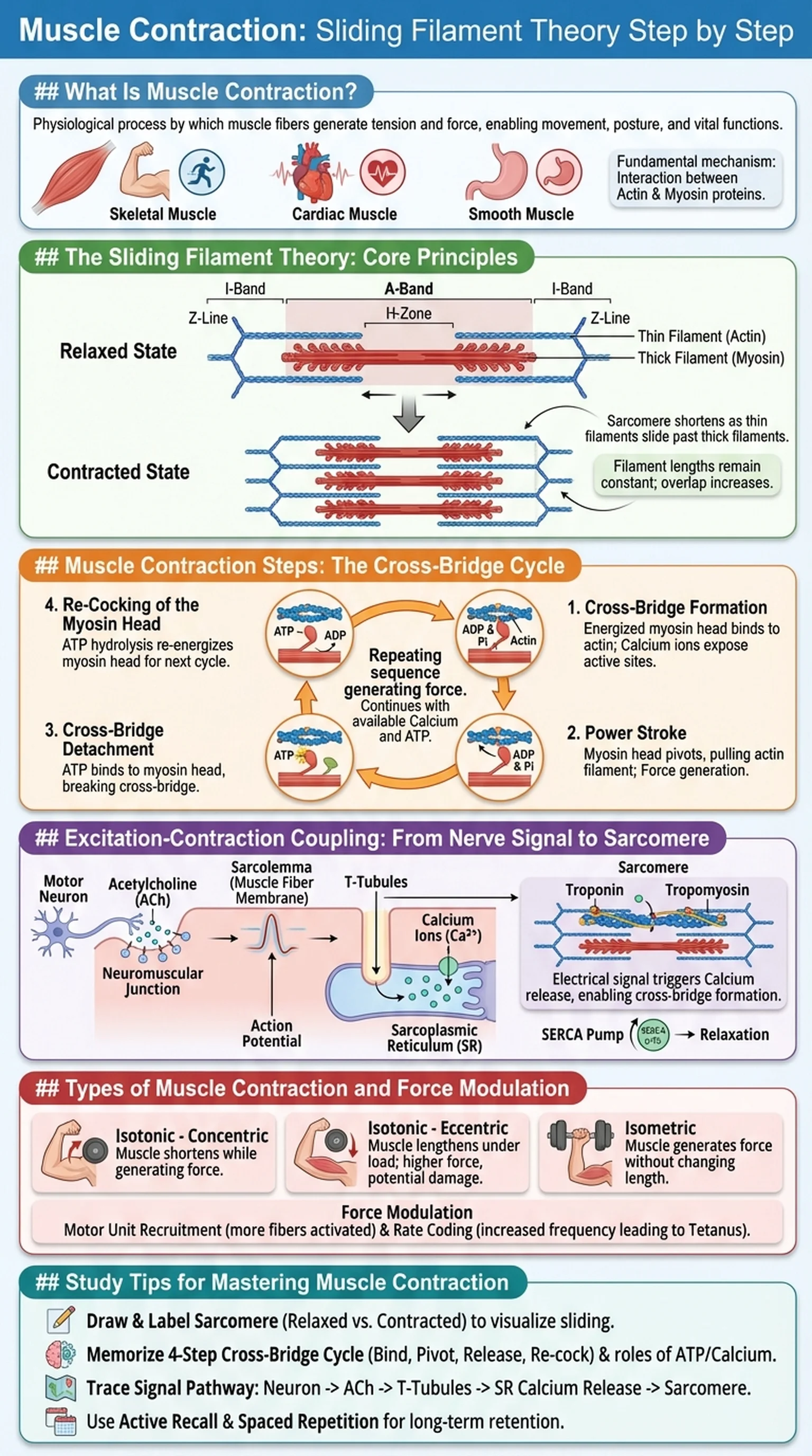
Muscle contraction is the physiological process by which muscle fibers generate tension and produce force, enabling movement, posture maintenance, and vital functions such as heartbeat and digestion. At the cellular level, muscle contraction involves the coordinated interaction of contractile proteins within specialized cells called muscle fibers, or myocytes. Whether you are lifting a weight, blinking an eye, or pumping blood through your circulatory system, muscle contraction is the fundamental mechanism at work.

There are three types of muscle tissue in the human body: skeletal, cardiac, and smooth. Skeletal muscle is under voluntary control and is responsible for locomotion and purposeful movements. Cardiac muscle contracts rhythmically to pump blood, and smooth muscle lines the walls of hollow organs and blood vessels, controlling involuntary functions like peristalsis. While the underlying molecular mechanism differs slightly among these types, the basic principle of muscle contraction remains rooted in the interaction between the proteins actin and myosin.

The study of muscle contraction is central to anatomy, physiology, kinesiology, and clinical medicine. Understanding the muscle contraction steps at the molecular level allows students to appreciate how defects in the contractile machinery lead to diseases such as muscular dystrophy, myasthenia gravis, and hypertrophic cardiomyopathy. Furthermore, the concept of the sarcomere as the functional unit of contraction provides an elegant framework for understanding how microscopic protein movements translate into macroscopic force generation. In this article, we will explore the sliding filament theory in detail, walking through each step of the cross-bridge cycle and explaining how the sarcomere shortens to produce muscle contraction.

Key Terms

Muscle Contraction

The physiological process by which muscle fibers generate tension through the interaction of contractile proteins, producing force and movement.

Sarcomere

The basic functional and structural unit of skeletal and cardiac muscle, defined as the segment between two Z-lines.

Actin

A thin filament protein in the sarcomere that provides binding sites for myosin heads during muscle contraction.

Myosin

A thick filament protein with globular heads that bind to actin and generate force through the power stroke during muscle contraction.

The Sliding Filament Theory: Core Principles

The sliding filament theory is the widely accepted model that explains how muscle contraction occurs at the molecular level. First proposed independently by Andrew Huxley and Rolf Niedergerke, and by Hugh Huxley and Jean Hanson in 1954, the sliding filament theory states that muscle shortening results from the sliding of thin filaments (actin) past thick filaments (myosin) within the sarcomere, without either filament actually changing in length. This elegant mechanism explains how microscopic protein interactions translate into the macroscopic shortening of an entire muscle.

According to the sliding filament theory, the sarcomere is the fundamental contractile unit. Each sarcomere is bounded by Z-lines (also called Z-discs), from which thin actin filaments extend toward the center. Thick myosin filaments occupy the central region of the sarcomere, overlapping with the actin filaments in a zone of interaction. When muscle contraction is triggered, the myosin heads reach out, bind to actin, and pull the thin filaments toward the center of the sarcomere. As the actin filaments slide inward, the Z-lines are drawn closer together, and the sarcomere shortens. Since thousands of sarcomeres are arranged in series along a myofibril, the cumulative shortening of each sarcomere results in the contraction of the entire muscle fiber.

A critical aspect of the sliding filament theory is that the lengths of the individual actin and myosin filaments remain constant during contraction. Only the degree of overlap between them changes. This was demonstrated through electron microscopy studies showing that the A-band (the region containing myosin) maintains a constant width, while the I-band (the region containing only actin) and the H-zone (the central region containing only myosin) both narrow during contraction. These observations provided the definitive structural evidence supporting the sliding filament theory and established it as the cornerstone of our understanding of muscle contraction.

Key Terms

Sliding Filament Theory

The model explaining muscle contraction as the result of actin thin filaments sliding past myosin thick filaments within the sarcomere, without filament length changes.

Z-Line (Z-Disc)

The boundary structure at each end of a sarcomere to which actin thin filaments are anchored.

A-Band

The dark band in a sarcomere that spans the entire length of the myosin thick filaments; its width remains constant during contraction.

I-Band

The light band in a sarcomere that contains only actin thin filaments; it shortens during muscle contraction.

H-Zone

The central region of the A-band containing only myosin thick filaments; it narrows during contraction as actin slides inward.

Muscle Contraction Steps: The Cross-Bridge Cycle

The muscle contraction steps describe the molecular events of the cross-bridge cycle, which is the repeating sequence of interactions between actin and myosin that produces force. Each cycle of cross-bridge formation, power stroke, detachment, and re-cocking of the myosin head constitutes one iteration of this process, and thousands of cycles occur simultaneously across a single sarcomere during contraction.

Step 1: Cross-Bridge Formation. The cycle begins when a myosin head, which has been energized by the hydrolysis of ATP into ADP and inorganic phosphate (Pi), binds to an exposed active site on the actin thin filament. This binding forms a cross-bridge between the thick and thin filaments. The myosin head can only bind when the regulatory proteins tropomyosin and troponin have shifted position, an event that requires calcium ions.

Step 2: Power Stroke. Once the cross-bridge is formed, the myosin head pivots, pulling the actin filament toward the center of the sarcomere. During this power stroke, ADP and Pi are released from the myosin head. The power stroke is the force-generating step of muscle contraction and is responsible for the sliding of actin past myosin as described by the sliding filament theory.

Step 3: Cross-Bridge Detachment. A new molecule of ATP binds to the myosin head, causing it to detach from actin. Without ATP, the myosin head remains locked onto actin, which is the molecular basis of rigor mortis after death.

Step 4: Re-Cocking of the Myosin Head. The myosin head hydrolyzes the newly bound ATP into ADP and Pi, using the released energy to return to its high-energy, cocked position. The myosin head is now ready to bind to a new site on actin further along the thin filament, and the cycle repeats. These muscle contraction steps continue as long as calcium is present and ATP is available, allowing sustained force generation within the sarcomere.

Key Terms

Cross-Bridge Cycle

The repeating sequence of myosin binding to actin, power stroke, detachment, and re-cocking that drives muscle contraction.

Power Stroke

The pivoting motion of the myosin head that pulls the actin filament toward the sarcomere center, generating force.

Tropomyosin

A regulatory protein that blocks myosin-binding sites on actin in resting muscle and shifts position when calcium binds to troponin.

Troponin

A regulatory protein complex on the thin filament that binds calcium ions, triggering the conformational change that exposes actin's active sites.

Rigor Mortis

The stiffening of muscles after death caused by the inability of myosin to detach from actin in the absence of ATP.

Excitation-Contraction Coupling: From Nerve Signal to Sarcomere Shortening

Excitation-contraction coupling is the sequence of events that links the electrical stimulation of a muscle fiber to the mechanical response of muscle contraction. This process bridges the gap between the nervous system's command and the sarcomere's execution of the sliding filament theory, ensuring that every nerve impulse translates into a coordinated contractile response.

The process begins at the neuromuscular junction, where a motor neuron releases the neurotransmitter acetylcholine (ACh) into the synaptic cleft. ACh binds to nicotinic receptors on the muscle fiber membrane (sarcolemma), triggering an action potential that propagates along the sarcolemma and deep into the muscle fiber through invaginations called T-tubules (transverse tubules). The T-tubules carry the electrical signal to the interior of the fiber, where they are closely associated with the sarcoplasmic reticulum (SR), a specialized endoplasmic reticulum that stores calcium ions.

When the action potential reaches the T-tubule-SR junction, voltage-sensitive receptors on the T-tubule (dihydropyridine receptors) undergo a conformational change that opens calcium release channels (ryanodine receptors) on the SR membrane. Calcium ions flood from the SR into the cytoplasm (sarcoplasm), rapidly increasing intracellular calcium concentration. Calcium binds to troponin C on the thin filaments within the sarcomere, causing a conformational shift in the troponin-tropomyosin complex. This shift moves tropomyosin away from the myosin-binding sites on actin, exposing them and allowing cross-bridge formation to begin.

Muscle contraction continues as long as calcium remains elevated in the sarcoplasm and ATP is available for the cross-bridge cycle. Relaxation occurs when calcium is actively pumped back into the sarcoplasmic reticulum by the SERCA (sarco-endoplasmic reticulum calcium ATPase) pump. As calcium levels drop, tropomyosin slides back over the actin binding sites, cross-bridges can no longer form, and the muscle fiber returns to its resting length. This entire process of excitation-contraction coupling ensures precise control over muscle contraction, allowing for graded responses ranging from gentle finger movements to maximal force production.

Key Terms

Excitation-Contraction Coupling

The sequence of events linking the electrical excitation of a muscle fiber to the mechanical response of contraction via calcium release.

T-Tubules

Transverse tubules that carry action potentials deep into the muscle fiber, connecting the sarcolemma to the sarcoplasmic reticulum.

Sarcoplasmic Reticulum

A specialized endoplasmic reticulum in muscle fibers that stores and releases calcium ions to regulate contraction.

Ryanodine Receptors

Calcium release channels on the sarcoplasmic reticulum membrane that open in response to signals from T-tubule receptors.

SERCA Pump

An ATP-dependent calcium pump that transports calcium back into the sarcoplasmic reticulum, enabling muscle relaxation.

Types of Muscle Contraction and Force Modulation

Muscle contraction can be classified into several types based on whether the muscle changes length and the direction of that change. Understanding these classifications is important for students of anatomy, kinesiology, and sports science, as different types of contraction serve distinct functional roles in movement and stability.

Isotonic contractions involve a change in muscle length while tension remains relatively constant. Isotonic contractions are further divided into concentric and eccentric types. In a concentric contraction, the muscle shortens as it generates force, such as the biceps brachii during the lifting phase of a curl. The actin myosin cross-bridges cycle in the manner described by the sliding filament theory, pulling the Z-lines closer together and shortening each sarcomere. In an eccentric contraction, the muscle lengthens while still generating tension, such as the biceps brachii during the lowering phase of a curl. Here, the external load exceeds the force generated by the cross-bridges, and the actin filaments slide outward even as myosin heads attempt to pull them inward. Eccentric contractions are associated with greater muscle damage and delayed-onset muscle soreness.

Isometric contractions occur when the muscle generates force without changing length. The actin myosin cross-bridges cycle and generate tension, but the external resistance exactly matches the internal force, so the sarcomere length and overall muscle length remain unchanged. Holding a heavy object at arm's length is an example of isometric contraction.

The force produced during muscle contraction is modulated by two primary mechanisms: motor unit recruitment and rate coding. Motor unit recruitment involves activating additional motor units to increase the total number of muscle fibers contracting simultaneously. Rate coding involves increasing the frequency of action potentials sent to a motor unit, leading to temporal summation and ultimately tetanus, a state of sustained maximal contraction. Together, these mechanisms allow the nervous system to finely control the muscle contraction steps and produce forces ranging from the delicate grip needed to hold a pen to the explosive power required for a vertical jump.

Key Terms

Concentric Contraction

A type of isotonic contraction in which the muscle shortens while generating force.

Eccentric Contraction

A type of isotonic contraction in which the muscle lengthens while generating force against a load that exceeds its contractile force.

Isometric Contraction

A contraction in which the muscle generates force without changing length, as the external load equals the internal force.

Motor Unit Recruitment

The activation of additional motor units to increase the force of muscle contraction.

Tetanus

A state of sustained maximal muscle contraction resulting from high-frequency stimulation that prevents relaxation between individual twitches.

Study Tips for Mastering Muscle Contraction

Muscle contraction and the sliding filament theory are among the most frequently tested topics in anatomy, physiology, and medical board examinations including the USMLE and MCAT. The topic requires integration of structural anatomy, protein biochemistry, neurophysiology, and cellular signaling, making it both challenging and rewarding to master.

First, learn the sarcomere structure inside and out. Draw a labeled diagram of a sarcomere showing the Z-lines, I-bands, A-band, H-zone, M-line, actin thin filaments, and myosin thick filaments. Then draw a second diagram showing the same sarcomere in a contracted state. Compare the two to verify your understanding of which bands shorten and which remain constant. This visual approach reinforces the core principle of the sliding filament theory: filaments slide, but their individual lengths do not change.

Second, memorize the muscle contraction steps of the cross-bridge cycle as a four-step loop: bind, pivot, release, re-cock. For each step, know which molecule (ATP, ADP, Pi, or calcium) is involved and what role it plays. A common exam question asks what happens when ATP is depleted (answer: rigor mortis, because myosin cannot detach from actin). Another frequently tested concept is the role of calcium in triggering muscle contraction by binding to troponin and exposing actin myosin binding sites.

Third, trace the entire signal pathway from nerve impulse to sarcomere shortening. Start at the motor neuron, follow acetylcholine across the neuromuscular junction, track the action potential along the sarcolemma and into the T-tubules, note the calcium release from the sarcoplasmic reticulum, and end at the cross-bridge cycle within the sarcomere. Understanding this complete sequence of excitation-contraction coupling is essential for answering integration-style questions.

Finally, use active recall and spaced repetition to cement your knowledge. Platforms like LectureScribe can generate flashcards and slide decks from your muscle contraction lecture notes, allowing you to quiz yourself on the sliding filament theory, actin myosin interactions, and muscle contraction steps until the material becomes second nature.

Key Terms

Active Recall

A study technique in which learners actively retrieve information from memory rather than passively reviewing notes.

Spaced Repetition

A learning strategy that involves reviewing material at increasing intervals to strengthen long-term memory retention.

MCAT

The Medical College Admission Test; a standardized exam required for admission to medical schools in the United States and Canada.

Frequently Asked Questions

What is the sliding filament theory?

The sliding filament theory explains muscle contraction as the result of actin thin filaments sliding past myosin thick filaments within the sarcomere. The filaments themselves do not change length; instead, myosin heads pull actin toward the center of the sarcomere, shortening it and generating force.

What are the steps of muscle contraction?

The muscle contraction steps follow the cross-bridge cycle: (1) cross-bridge formation when energized myosin binds to actin, (2) power stroke as myosin pivots and pulls actin, (3) detachment when new ATP binds to myosin, and (4) re-cocking as myosin hydrolyzes ATP and returns to its high-energy position.

What role do actin and myosin play in muscle contraction?

Actin forms the thin filaments that provide binding sites for myosin. Myosin forms the thick filaments with globular heads that bind to actin, perform the power stroke to generate force, and drive the sliding of filaments within the sarcomere. The actin myosin interaction is the molecular basis of all muscle contraction.

What is a sarcomere?

A sarcomere is the basic functional unit of skeletal and cardiac muscle, defined as the segment between two Z-lines. It contains organized arrays of actin thin filaments and myosin thick filaments. Shortening of the sarcomere through the sliding filament mechanism produces muscle contraction.

Why is calcium important for muscle contraction?

Calcium ions are essential for initiating muscle contraction. When calcium is released from the sarcoplasmic reticulum, it binds to troponin on the actin filament, causing tropomyosin to shift and expose myosin-binding sites. Without calcium, the binding sites remain blocked and cross-bridge cycling cannot occur.

What causes rigor mortis?

Rigor mortis occurs after death when ATP is depleted. Without ATP, myosin heads cannot detach from actin after the power stroke, leaving the cross-bridges permanently locked. This results in the characteristic stiffness of muscles following death.

How does the sliding filament theory explain the shortening of a sarcomere?

According to the sliding filament theory, myosin heads bind to actin and pull the thin filaments toward the center of the sarcomere. As actin slides inward, the Z-lines are pulled closer together, shortening the sarcomere. The I-band and H-zone narrow, while the A-band stays constant, because the filaments slide rather than shrink.

Study Anatomy Smarter

Upload your lecture notes and get AI-generated flashcards, quizzes, infographics, and study guides tailored to your curriculum.

Try LectureScribe Free

Create Study Materials

Transform your Anatomy lectures into flashcards, quizzes, and visual study guides with AI.

  • AI-generated flashcards & quizzes
  • Visual infographics from notes
  • Interactive study shorts
Start Studying Free

Quick Facts

SubjectAnatomy
Word Count2,500+
Slides5
InfographicIncluded
AuthorLectureScribe

Master Anatomy with LectureScribe

Upload your lecture notes and get AI-powered flashcards, quizzes, infographics, and study guides in minutes.