LectureScribe Logo
lecturescribe.io

LOCK IN VALENTINE PRICING BEFORE SALE ENDS!

Use code — New joiners get 40% OFF on annual plans

Plans will have higher prices after this sale ends

0days
:
0hours
:
0minutes
:
0seconds
Anatomy2,500+ words5 slides
🧠

Brain Anatomy: Lobes, Functions & Structures Guide

Learn brain anatomy including the brain lobes, brain functions, cerebral cortex, and key brain structures. A comprehensive guide to understanding the human brain.

L
LectureScribeAI-Powered Study Platform

Study Infographic

Complete brain anatomy diagram illustrating the brain lobes (frontal, parietal, temporal, occipital), cerebral cortex functional areas, and key brain structures.

Download
Brain anatomy diagram showing the four brain lobes, cerebral cortex, and major brain structures with their functions labeled

Interactive Study Short

Swipe through 5 slides about Brain Anatomy: Lobes, Functions & Structures Guide

1 / 5

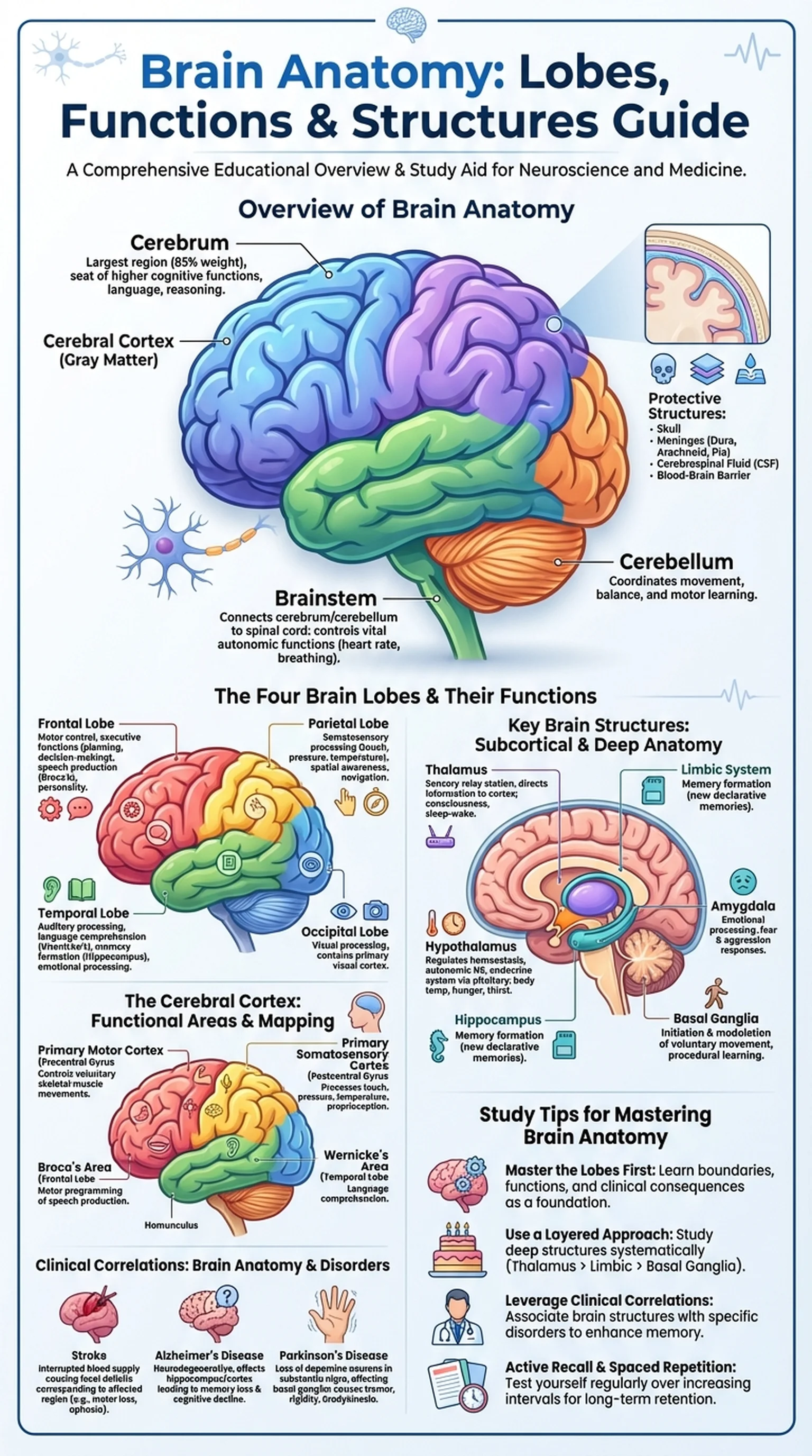
Overview of Brain Anatomy

Brain anatomy is the study of the structural organization of the human brain, the most complex organ in the body. Weighing approximately 1.4 kilograms and containing roughly 86 billion neurons, the brain is the command center of the nervous system, responsible for controlling thought, memory, emotion, movement, sensation, and virtually every process that regulates the body. Understanding brain anatomy is fundamental for students of neuroscience, anatomy, psychology, and medicine.

The brain can be broadly divided into three major regions: the cerebrum, the cerebellum, and the brainstem. The cerebrum is the largest portion, accounting for about 85% of the brain's total weight. Its outer surface, the cerebral cortex, is a highly folded layer of gray matter that is the seat of higher cognitive functions including language, reasoning, perception, and voluntary movement. The cerebellum, located at the posterior base of the brain, coordinates movement, balance, and motor learning. The brainstem, consisting of the midbrain, pons, and medulla oblongata, connects the cerebrum and cerebellum to the spinal cord and controls vital autonomic functions such as heart rate, breathing, and blood pressure.

The brain is protected by multiple layers of defense. The skull (cranium) provides rigid bony protection. Beneath the skull, three layers of connective tissue membranes called meninges (dura mater, arachnoid mater, and pia mater) surround and cushion the brain. Cerebrospinal fluid (CSF), produced by the choroid plexus in the brain's ventricles, circulates around and within the brain, providing buoyancy, nutrient delivery, and waste removal. The blood-brain barrier, formed by tight junctions between endothelial cells of brain capillaries, selectively controls which substances can enter the brain from the bloodstream. Together, these protective brain structures ensure the delicate neural tissue is shielded from mechanical injury, infection, and toxic substances.

Key Terms

Brain Anatomy

The study of the structural organization of the brain, including its major divisions, brain lobes, cortical regions, and subcortical brain structures.

Cerebral Cortex

The outer layer of gray matter covering the cerebrum; responsible for higher cognitive functions including perception, thought, language, and voluntary movement.

Cerebellum

A brain structure located at the posterior base of the skull that coordinates voluntary movement, balance, posture, and motor learning.

Brainstem

The region of the brain connecting the cerebrum and cerebellum to the spinal cord, comprising the midbrain, pons, and medulla oblongata.

Cerebrospinal Fluid (CSF)

A clear fluid produced by the choroid plexus that circulates within the ventricles and around the brain, providing cushioning and nutrient transport.

The Four Brain Lobes and Their Functions

The cerebral cortex of each hemisphere is divided into four brain lobes, each named after the overlying cranial bone: the frontal lobe, parietal lobe, temporal lobe, and occipital lobe. Each of these brain lobes is associated with specific brain functions, although it is important to recognize that most complex behaviors involve the coordinated activity of multiple lobes working together.

The frontal lobe is the largest of the brain lobes, occupying the anterior portion of each hemisphere. It is separated from the parietal lobe by the central sulcus and from the temporal lobe by the lateral (Sylvian) fissure. The frontal lobe is responsible for a wide range of brain functions, including voluntary motor control (via the primary motor cortex in the precentral gyrus), executive functions such as planning, decision-making, and impulse control (via the prefrontal cortex), speech production (via Broca's area in the left hemisphere), and personality expression. Damage to the frontal lobe can result in personality changes, impaired judgment, motor deficits, and expressive aphasia.

The parietal lobe is located posterior to the central sulcus and superior to the temporal lobe. Its primary function is the processing of somatosensory information, including touch, pressure, temperature, and proprioception. The primary somatosensory cortex, located in the postcentral gyrus, receives sensory input from the contralateral side of the body. The parietal lobe also plays important roles in spatial awareness, navigation, and the integration of sensory information from multiple modalities.

The temporal lobe is situated on the lateral side of each hemisphere, below the lateral fissure. It is critical for auditory processing (via the primary auditory cortex), language comprehension (via Wernicke's area in the dominant hemisphere), memory formation (in association with the hippocampus), and emotional processing (in association with the amygdala). The occipital lobe is the most posterior of the brain lobes and is primarily dedicated to visual processing. The primary visual cortex (area V1) receives input from the retina via the lateral geniculate nucleus of the thalamus and performs the initial cortical processing of visual information. Understanding the brain lobes and their associated brain functions is a cornerstone of neuroanatomy and clinical neuroscience.

Key Terms

Brain Lobes

The four major divisions of the cerebral cortex: frontal, parietal, temporal, and occipital lobes, each associated with specific brain functions.

Frontal Lobe

The anterior brain lobe responsible for motor control, executive functions, speech production, and personality.

Parietal Lobe

The brain lobe posterior to the central sulcus that processes somatosensory information including touch, temperature, and proprioception.

Temporal Lobe

The lateral brain lobe involved in auditory processing, language comprehension, memory formation, and emotional processing.

Occipital Lobe

The posterior brain lobe primarily dedicated to visual processing, containing the primary visual cortex.

Key Brain Structures: Subcortical and Deep Anatomy

Beneath the cerebral cortex lies a collection of critical brain structures that play essential roles in emotion, memory, movement, sensory relay, and homeostasis. These subcortical and deep brain structures work in concert with the cerebral cortex to produce the full spectrum of brain functions that define human behavior and physiology.

The thalamus is a paired, egg-shaped structure located at the center of the brain. Often called the "relay station" of the brain, the thalamus receives sensory information from virtually all sensory systems (except olfaction) and routes it to the appropriate areas of the cerebral cortex for processing. The thalamus also plays roles in consciousness, sleep-wake regulation, and attention. Damage to the thalamus can produce widespread sensory deficits and altered consciousness.

The hypothalamus, located just below the thalamus, is a small but vital brain structure that serves as the primary link between the nervous system and the endocrine system. Despite its small size, the hypothalamus controls body temperature, hunger, thirst, circadian rhythms, and the autonomic nervous system. It exerts its endocrine influence by regulating the pituitary gland, which in turn controls the release of hormones throughout the body.

The limbic system is a group of interconnected brain structures involved in emotion, motivation, and memory. Key components include the hippocampus (essential for the formation of new declarative memories), the amygdala (central to emotional processing, particularly fear and aggression), and the cingulate gyrus (involved in emotional regulation and pain processing). The basal ganglia, a group of nuclei deep within the cerebral hemispheres, are critical for the initiation and modulation of voluntary movement. The major components of the basal ganglia include the caudate nucleus, putamen, and globus pallidus. Dysfunction of the basal ganglia is implicated in movement disorders such as Parkinson's disease (characterized by tremor, rigidity, and bradykinesia due to dopamine depletion) and Huntington's disease (characterized by involuntary choreiform movements due to degeneration of the caudate nucleus). Together, these brain structures form the deep architecture upon which the higher cortical brain functions depend.

Key Terms

Thalamus

A paired brain structure that acts as the main sensory relay center, directing incoming sensory information to the appropriate regions of the cerebral cortex.

Hypothalamus

A small brain structure below the thalamus that regulates homeostasis, the autonomic nervous system, and the endocrine system via the pituitary gland.

Hippocampus

A limbic brain structure essential for the consolidation of short-term memories into long-term declarative memories.

Amygdala

A limbic brain structure central to emotional processing, particularly the generation and regulation of fear responses.

Basal Ganglia

A group of subcortical nuclei involved in the regulation of voluntary movement, procedural learning, and habit formation.

The Cerebral Cortex: Functional Areas and Mapping

The cerebral cortex is the thin, folded outer layer of the cerebrum that is responsible for the highest levels of brain functions, including conscious perception, thought, language, and voluntary action. The cortex is composed of gray matter, meaning it is dense with neuronal cell bodies, dendrites, and synapses. The extensive folding of the cerebral cortex into gyri (ridges) and sulci (grooves) dramatically increases its surface area, allowing approximately 16 billion neurons to be packed into a structure only 2-4 millimeters thick.

Functional areas of the cerebral cortex can be classified into three broad categories: primary cortical areas, association areas, and multimodal integration areas. Primary cortical areas are directly involved in sensory processing or motor output. The primary motor cortex (precentral gyrus, frontal lobe) controls voluntary skeletal muscle movements and is organized somatotopically, meaning different regions control different body parts, as depicted in the motor homunculus. The primary somatosensory cortex (postcentral gyrus, parietal lobe) processes tactile and proprioceptive input and is similarly organized. The primary visual cortex is in the occipital lobe, and the primary auditory cortex is in the temporal lobe.

Association areas surround the primary areas and are responsible for higher-order processing. The prefrontal association area, in the anterior frontal lobe, is involved in personality, executive function, working memory, and moral reasoning. The parietal association areas integrate sensory information to create spatial awareness and body schema. Wernicke's area, in the posterior temporal lobe of the dominant hemisphere, is critical for language comprehension. Broca's area, in the inferior frontal lobe, controls the motor programming of speech.

The cerebral cortex also contains multimodal association areas where information from multiple senses converges. These regions, including the posterior parietal cortex and the prefrontal cortex, enable complex brain functions such as abstract thinking, planning, and self-awareness. The study of cortical mapping has been advanced by techniques such as functional MRI (fMRI) and positron emission tomography (PET), which allow researchers to visualize brain activity in living subjects and identify the brain anatomy underlying specific cognitive tasks.

Key Terms

Primary Motor Cortex

The region of the cerebral cortex in the precentral gyrus that directly controls voluntary skeletal muscle movements.

Primary Somatosensory Cortex

The region of the cerebral cortex in the postcentral gyrus that processes touch, pressure, temperature, and proprioceptive input.

Broca's Area

A region in the inferior frontal lobe of the dominant hemisphere responsible for the motor programming of speech production.

Wernicke's Area

A region in the posterior temporal lobe of the dominant hemisphere critical for language comprehension.

Homunculus

A distorted figure representing the somatotopic mapping of the body on the primary motor or somatosensory cortex of the cerebral cortex.

Clinical Correlations: Brain Anatomy and Neurological Disorders

A detailed understanding of brain anatomy is essential for diagnosing and managing neurological disorders, as the location of a lesion within the brain determines the specific symptoms a patient will exhibit. The principle of structure-function correlation is the foundation of clinical neurology: by identifying which brain functions are impaired, clinicians can predict which brain structures or brain lobes are affected, and vice versa.

Stroke is one of the most common neurological emergencies and vividly illustrates the clinical importance of brain anatomy. An ischemic stroke in the territory of the middle cerebral artery, which supplies the lateral cerebral cortex, can produce contralateral motor and sensory deficits (from damage to the primary motor and somatosensory cortices), expressive aphasia (from damage to Broca's area in the frontal lobe), or receptive aphasia (from damage to Wernicke's area in the temporal lobe). A stroke affecting the posterior cerebral artery can cause visual field deficits due to damage to the occipital lobe. Knowledge of the vascular territories and their relationship to brain lobes allows clinicians to rapidly localize the lesion and initiate time-critical treatment.

Neurodegenerative diseases also demonstrate the connection between brain structures and brain functions. Alzheimer's disease, the most common cause of dementia, initially affects the hippocampus and entorhinal cortex, leading to progressive memory loss. As the disease advances, it spreads to the cerebral cortex, impairing language, reasoning, and visuospatial abilities. Parkinson's disease targets dopaminergic neurons in the substantia nigra of the midbrain, leading to motor symptoms such as tremor, rigidity, and bradykinesia due to disrupted basal ganglia circuits.

Traumatic brain injury (TBI) provides further clinical illustration. A blow to the frontal region may damage the prefrontal cortex, resulting in personality changes and impaired executive function. Temporal lobe injuries can cause memory deficits and seizures. Understanding brain anatomy at this level of detail allows healthcare providers to predict outcomes, guide rehabilitation, and counsel patients and families about the expected course of recovery.

Key Terms

Stroke

A neurological emergency caused by interrupted blood supply to the brain, resulting in focal deficits that correspond to the affected brain regions.

Aphasia

An impairment of language ability caused by damage to the language areas of the cerebral cortex, including Broca's area (expressive) or Wernicke's area (receptive).

Alzheimer's Disease

A progressive neurodegenerative disorder that initially affects the hippocampus and cerebral cortex, leading to memory loss and cognitive decline.

Parkinson's Disease

A neurodegenerative movement disorder caused by the loss of dopaminergic neurons in the substantia nigra, leading to tremor, rigidity, and bradykinesia.

Study Tips for Mastering Brain Anatomy

Brain anatomy is one of the most challenging and most frequently tested topics in anatomy, neuroscience, and medical board examinations such as the USMLE and MCAT. The sheer number of brain structures, their interconnections, and their associated brain functions can feel overwhelming without a structured study approach. Here are evidence-based strategies for learning brain anatomy effectively.

First, learn the brain lobes and their primary functions as your foundation. Before diving into subcortical structures and pathways, make sure you can identify the frontal, parietal, temporal, and occipital lobes on a lateral, medial, and inferior view of the brain. For each lobe, know its boundaries (what sulci and fissures define it), its primary functional areas, and the clinical consequences of damage to that lobe. This lobar framework serves as a scaffold onto which all other details of brain anatomy can be attached.

Second, use a layered approach to learn deep brain structures. Start with the thalamus and hypothalamus, then move to the limbic structures (hippocampus, amygdala, cingulate gyrus), and then the basal ganglia. For each structure, learn its location, primary function, major connections, and associated clinical conditions. Creating a table with columns for structure, function, and clinical significance is an efficient way to organize this information. Knowing the brain structures in this systematic way is far more effective than trying to learn them in random order.

Third, leverage clinical correlations. For every brain region you study, ask yourself: what happens when this area is damaged? The clinical significance of brain anatomy is not only tested heavily on exams but also makes the material more engaging and memorable. For instance, associating the cerebral cortex with cortical strokes, the hippocampus with Alzheimer's disease, and the basal ganglia with Parkinson's disease creates lasting memory anchors.

Finally, use active recall and spaced repetition to consolidate your knowledge over time. Platforms like LectureScribe can generate flashcards, slide decks, and practice questions from your brain anatomy lecture notes, allowing you to test yourself repeatedly on brain lobes, brain functions, and brain structures. Combining visual study (labeling diagrams) with self-testing is the most effective way to achieve mastery of this essential anatomical topic.

Key Terms

Active Recall

A study technique in which learners actively retrieve information from memory rather than passively reviewing notes.

Spaced Repetition

A learning strategy that involves reviewing material at increasing intervals to strengthen long-term memory retention.

MCAT

The Medical College Admission Test; a standardized exam required for admission to medical schools in the United States and Canada.

Frequently Asked Questions

What are the four brain lobes and their functions?

The four brain lobes are the frontal lobe (motor control, executive functions, speech), parietal lobe (somatosensory processing, spatial awareness), temporal lobe (auditory processing, language comprehension, memory), and occipital lobe (visual processing). Each brain lobe contributes specific brain functions while working together for complex behaviors.

What is the cerebral cortex?

The cerebral cortex is the thin, folded outer layer of gray matter covering the cerebrum. It is 2-4 mm thick and contains approximately 16 billion neurons. The cerebral cortex is responsible for higher brain functions including conscious perception, thought, language, memory, and voluntary movement.

What are the major brain structures?

Major brain structures include the cerebrum (with its cerebral cortex and four brain lobes), cerebellum, brainstem, thalamus, hypothalamus, hippocampus, amygdala, and basal ganglia. Each of these brain structures performs specific functions essential for normal brain activity and behavior.

What does the frontal lobe do?

The frontal lobe controls voluntary motor movement (via the primary motor cortex), executive functions such as planning and decision-making (via the prefrontal cortex), speech production (via Broca's area), and personality expression. It is the largest of the brain lobes and is critical for higher-order brain functions.

What is the role of the hippocampus in brain anatomy?

The hippocampus is a limbic brain structure essential for converting short-term memories into long-term declarative memories. It is located in the medial temporal lobe and is one of the first brain structures affected in Alzheimer's disease, which is why memory loss is an early symptom.

How does brain anatomy relate to neurological disorders?

Brain anatomy directly determines the symptoms of neurological disorders. Damage to specific brain lobes or brain structures produces predictable deficits: frontal lobe damage causes personality changes and motor deficits, temporal lobe damage impairs memory and language, and basal ganglia dysfunction causes movement disorders like Parkinson's disease.

What is the difference between gray matter and white matter in brain anatomy?

Gray matter, found in the cerebral cortex and deep brain nuclei, consists of neuronal cell bodies, dendrites, and synapses where information processing occurs. White matter, found beneath the cortex, consists of myelinated axon bundles that connect different brain structures and brain lobes, enabling communication across regions.

Study Anatomy Smarter

Upload your lecture notes and get AI-generated flashcards, quizzes, infographics, and study guides tailored to your curriculum.

Try LectureScribe Free

Create Study Materials

Transform your Anatomy lectures into flashcards, quizzes, and visual study guides with AI.

  • AI-generated flashcards & quizzes
  • Visual infographics from notes
  • Interactive study shorts
Start Studying Free

Quick Facts

SubjectAnatomy
Word Count2,500+
Slides5
InfographicIncluded
AuthorLectureScribe

Master Anatomy with LectureScribe

Upload your lecture notes and get AI-powered flashcards, quizzes, infographics, and study guides in minutes.