LectureScribe Logo
lecturescribe.io

LOCK IN VALENTINE PRICING BEFORE SALE ENDS!

Use code — New joiners get 40% OFF on annual plans

Plans will have higher prices after this sale ends

0days
:
0hours
:
0minutes
:
0seconds
Anatomy2,500+ words5 slides
🫁

Nephron Structure and Function: Kidney Filtration Explained

Learn nephron structure and function in detail. Understand how the nephron performs glomerular filtration, tubular reabsorption, and secretion to maintain kidney function and renal physiology.

L
LectureScribeAI-Powered Study Platform

Study Infographic

Complete nephron anatomy illustration showing each structural segment, key transporters, hormonal regulation sites, and the pathway of filtrate from glomerular filtration to urine production.

Download
Nephron structure diagram showing the glomerulus, Bowman's capsule, proximal convoluted tubule, loop of Henle, distal convoluted tubule, and collecting duct with labeled transport processes

Interactive Study Short

Swipe through 5 slides about Nephron Structure and Function: Kidney Filtration Explained

1 / 5

Introduction to the Nephron and Kidney Function

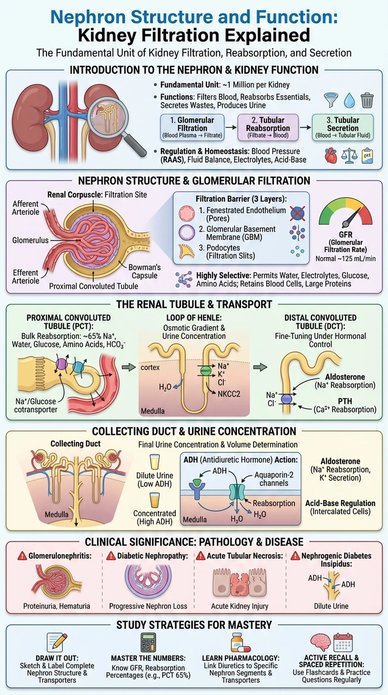
The nephron is the fundamental structural and functional unit of the kidney. Each human kidney contains approximately one million nephrons, and together they perform the critical tasks of filtering blood, reabsorbing essential substances, secreting waste products, and producing urine. Understanding nephron structure and nephron function is foundational for students of anatomy, physiology, and medicine because the kidneys regulate fluid balance, electrolyte concentrations, blood pressure, acid-base homeostasis, and the excretion of metabolic waste.

Kidney function depends on three sequential processes carried out by the nephron: glomerular filtration, tubular reabsorption, and tubular secretion. Glomerular filtration is the first step, in which blood plasma is filtered through the glomerular capillaries into Bowman's capsule. Tubular reabsorption recovers water, glucose, amino acids, and ions from the filtrate back into the peritubular capillaries. Tubular secretion adds additional waste products and excess ions from the blood into the tubular fluid. The final product, urine, represents only about 1% of the original filtrate volume, demonstrating the extraordinary efficiency of the nephron.

Renal physiology encompasses the study of how these processes are regulated to maintain homeostasis under varying conditions. When blood pressure drops, the kidneys activate the renin-angiotensin-aldosterone system (RAAS) to conserve sodium and water. When blood pressure rises, natriuretic peptides promote sodium and water excretion. The nephron is therefore not merely a passive filter but an active, hormonally responsive organ that adjusts its function moment by moment. A thorough understanding of nephron structure provides the anatomical framework necessary for comprehending these dynamic physiological and pathological processes.

Key Terms

Nephron

The fundamental structural and functional unit of the kidney, responsible for filtering blood, reabsorbing essential substances, secreting wastes, and producing urine.

Kidney Function

The collective physiological activities of the kidneys, including filtration, reabsorption, secretion, electrolyte balance, blood pressure regulation, and acid-base homeostasis.

Renal Physiology

The branch of physiology concerned with the function of the kidneys, including glomerular filtration, tubular transport, urine concentration, and hormonal regulation.

Glomerular Filtration

The process by which blood plasma is filtered through the glomerular capillaries and Bowman's capsule, producing a protein-free filtrate that enters the renal tubules.

Nephron Structure: Anatomy of the Renal Corpuscle

The nephron structure consists of two major components: the renal corpuscle, where filtration occurs, and the renal tubule, where reabsorption and secretion take place. The renal corpuscle is located in the kidney's cortex and is composed of the glomerulus (a tuft of fenestrated capillaries) and Bowman's capsule (a cup-shaped epithelial structure that surrounds the glomerulus). Blood enters the glomerulus through the afferent arteriole and exits through the efferent arteriole, a unique arrangement in which a capillary bed is sandwiched between two arterioles rather than between an arteriole and a venule.

The filtration barrier of the glomerulus has three layers that determine what passes from the blood into Bowman's capsule. The first layer is the fenestrated endothelium of the glomerular capillaries, which contains pores that allow passage of water and small solutes but retain blood cells. The second layer is the glomerular basement membrane (GBM), a dense meshwork of collagen and glycoproteins that blocks most proteins by size and charge. The third layer is the visceral epithelium of Bowman's capsule, composed of specialized cells called podocytes, which extend foot processes (pedicels) that interdigitate to form filtration slits. Together, these three layers create a highly selective barrier that permits glomerular filtration of water, electrolytes, glucose, amino acids, and small waste molecules while retaining blood cells and large plasma proteins.

The glomerular filtration rate (GFR) is the volume of filtrate produced per unit time and is a key clinical measure of kidney function. Normal GFR is approximately 125 mL/min or 180 liters per day. It is determined by the net filtration pressure, which depends on the balance of hydrostatic and oncotic pressures across the glomerular capillary wall. Changes in afferent or efferent arteriolar tone, blood pressure, or plasma protein concentration all affect GFR, making the renal corpuscle a highly regulated component of nephron structure.

Key Terms

Nephron Structure

The anatomical organization of the nephron, consisting of the renal corpuscle (glomerulus and Bowman's capsule) and the renal tubule (PCT, loop of Henle, DCT, and collecting duct).

Glomerulus

A tuft of fenestrated capillaries within Bowman's capsule where blood plasma is filtered to form the initial tubular fluid.

Podocytes

Specialized epithelial cells of Bowman's visceral layer whose foot processes form filtration slits, constituting the final barrier in glomerular filtration.

Glomerular Filtration Rate (GFR)

The volume of plasma filtered by the glomeruli per unit time (normally ~125 mL/min), used as a primary clinical indicator of kidney function.

Bowman's Capsule

The double-walled cup-shaped structure surrounding the glomerulus that collects the filtrate and channels it into the proximal convoluted tubule.

The Renal Tubule: Proximal Tubule, Loop of Henle, and Distal Tubule

After filtrate is formed in the renal corpuscle, it enters the renal tubule, a continuous series of segments that modify the filtrate's composition through reabsorption and secretion. The tubule begins with the proximal convoluted tubule (PCT), continues as the loop of Henle, transitions into the distal convoluted tubule (DCT), and finally empties into the collecting duct. Each segment of the nephron structure has distinct cellular characteristics and transport functions that contribute to the nephron's overall role in kidney function.

The proximal convoluted tubule is responsible for the bulk of reabsorption. Approximately 65% of filtered sodium, water, bicarbonate, glucose, and amino acids are reclaimed in the PCT. The cells lining the PCT have a prominent brush border of microvilli that dramatically increases surface area and abundant mitochondria that power active transport via Na+/K+-ATPase pumps on the basolateral membrane. Glucose and amino acids are reabsorbed by sodium-dependent cotransporters on the apical membrane. The PCT also secretes organic acids, organic bases, and certain drugs into the tubular fluid.

The loop of Henle extends from the cortex into the medulla and back, and its primary role is to establish the osmotic gradient in the renal medulla that enables urine concentration. The thin descending limb is highly permeable to water but relatively impermeable to solutes, so water is reabsorbed by osmosis as the filtrate descends into the hyperosmotic medulla. The thick ascending limb is impermeable to water but actively transports sodium, potassium, and chloride out of the tubule via the Na+/K+/2Cl- cotransporter (NKCC2), diluting the tubular fluid and contributing to the medullary concentration gradient. The distal convoluted tubule fine-tunes sodium and calcium reabsorption under hormonal control. The Na+/Cl- cotransporter in the DCT reabsorbs sodium, and parathyroid hormone stimulates calcium reabsorption here. This segment is a key site for nephron function in maintaining electrolyte balance.

Key Terms

Proximal Convoluted Tubule (PCT)

The first segment of the renal tubule, responsible for reabsorbing approximately 65% of filtered sodium, water, glucose, amino acids, and bicarbonate.

Loop of Henle

The U-shaped tubular segment that establishes the medullary osmotic gradient, enabling the kidney to produce concentrated or dilute urine.

Distal Convoluted Tubule (DCT)

The tubular segment between the loop of Henle and the collecting duct, responsible for fine-tuning sodium and calcium reabsorption under hormonal regulation.

Na+/K+/2Cl- Cotransporter (NKCC2)

The apical membrane transporter in the thick ascending limb that reabsorbs sodium, potassium, and chloride; the target of loop diuretics such as furosemide.

The Collecting Duct and Urine Concentration

The collecting duct is the final segment of the nephron's tubular system and plays a decisive role in determining the final concentration and volume of urine. Although not technically part of the nephron in the strictest anatomical sense, the collecting duct is functionally inseparable from nephron function because it receives filtrate from multiple nephrons and makes the final adjustments that determine whether dilute or concentrated urine is produced. The collecting duct traverses the renal cortex and medulla, passing through progressively more hyperosmotic regions created by the countercurrent multiplier system of the loop of Henle.

The water permeability of the collecting duct is regulated by antidiuretic hormone (ADH, also called vasopressin), which is released from the posterior pituitary gland in response to increased plasma osmolality or decreased blood volume. ADH binds to V2 receptors on the basolateral membrane of collecting duct principal cells, triggering the insertion of aquaporin-2 water channels into the apical membrane. When ADH levels are high, the collecting duct becomes highly permeable to water, and water is reabsorbed by osmosis into the hypertonic medullary interstitium, producing small volumes of concentrated urine. When ADH levels are low, as after drinking large amounts of water, the collecting duct remains relatively impermeable to water, and dilute urine is excreted.

The collecting duct also contains intercalated cells that are critical for acid-base regulation. Type A intercalated cells secrete hydrogen ions and reabsorb bicarbonate, helping to correct metabolic acidosis. Type B intercalated cells secrete bicarbonate and reabsorb hydrogen ions during alkalosis. Additionally, aldosterone acts on principal cells of the collecting duct to stimulate sodium reabsorption and potassium secretion via the epithelial sodium channel (ENaC), linking nephron function to blood pressure regulation through the renin-angiotensin-aldosterone system. The collecting duct thus serves as the final checkpoint where renal physiology integrates hormonal signals to produce urine of appropriate composition and volume.

Key Terms

Collecting Duct

The terminal tubular segment that receives filtrate from multiple nephrons and determines final urine concentration under the control of ADH and aldosterone.

Antidiuretic Hormone (ADH)

A posterior pituitary hormone that increases water permeability of the collecting duct by promoting insertion of aquaporin-2 channels, concentrating the urine.

Aquaporin-2

Water channel proteins inserted into the apical membrane of collecting duct principal cells in response to ADH, enabling water reabsorption.

Countercurrent Multiplier

The mechanism by which the loop of Henle creates an increasing osmotic gradient in the renal medulla, enabling the production of concentrated urine.

Clinical Significance: Nephron Pathology and Kidney Disease

Damage to any component of the nephron structure can impair kidney function and lead to renal disease. Clinicians assess nephron function primarily through the glomerular filtration rate (GFR), serum creatinine levels, urinalysis, and urine electrolyte measurements. Understanding the anatomical basis of common kidney diseases helps connect the principles of renal physiology to clinical practice.

Glomerulonephritis refers to a group of diseases that damage the glomerular filtration barrier, allowing proteins and red blood cells to leak into the urine (proteinuria and hematuria). In minimal change disease, podocyte foot process effacement impairs the filtration slit mechanism, causing nephrotic syndrome with massive proteinuria. In IgA nephropathy, immune complex deposition in the glomerular mesangium triggers inflammation and glomerular filtration impairment. Diabetic nephropathy, the leading cause of chronic kidney disease worldwide, involves thickening of the glomerular basement membrane and expansion of the mesangial matrix, progressively reducing the number of functional nephrons.

Tubular and interstitial diseases also compromise nephron function. Acute tubular necrosis (ATN), caused by ischemia or nephrotoxins, damages the epithelial cells of the proximal tubule and the thick ascending limb, leading to acute kidney injury. Bartter syndrome and Gitelman syndrome are genetic disorders affecting the NKCC2 transporter in the thick ascending limb and the Na+/Cl- cotransporter in the DCT, respectively, causing electrolyte imbalances that mimic the effects of diuretics. Nephrogenic diabetes insipidus results from collecting duct resistance to ADH, producing large volumes of dilute urine despite adequate hormone levels. Each of these conditions illustrates how specific disruptions in nephron structure or function lead to predictable clinical syndromes, reinforcing the importance of a thorough understanding of renal physiology for clinical medicine.

Key Terms

Glomerulonephritis

A group of inflammatory diseases affecting the glomeruli that impair glomerular filtration and can cause proteinuria, hematuria, and progressive kidney damage.

Diabetic Nephropathy

Chronic kidney disease caused by diabetes mellitus, characterized by thickening of the glomerular basement membrane and progressive loss of nephron function.

Acute Tubular Necrosis

Death of tubular epithelial cells due to ischemia or toxins, causing acute kidney injury with impaired reabsorption and secretion.

Nephrogenic Diabetes Insipidus

A condition in which the collecting duct fails to respond to ADH, resulting in the excretion of large volumes of dilute urine.

Study Strategies for Nephron Anatomy and Renal Physiology

The nephron and kidney function are consistently high-yield topics on the MCAT, USMLE Step 1, and medical school physiology exams. Questions often require you to integrate anatomical knowledge of nephron structure with physiological principles of glomerular filtration, tubular transport, and hormonal regulation. Here are targeted strategies for mastering this material.

First, draw the nephron from memory. Sketch the complete nephron structure from the glomerulus through the collecting duct, labeling each segment: Bowman's capsule, PCT, descending thin limb, ascending thin limb, thick ascending limb, DCT, and collecting duct. At each segment, annotate the major substances reabsorbed and secreted, the transporters involved, and the hormones that regulate transport. This exercise transforms the nephron from an abstract concept into a concrete spatial map that you can mentally traverse during exams.

Second, master the numbers. Know that GFR is approximately 125 mL/min, that the PCT reabsorbs about 65% of filtered sodium and water, and that only 1% of the original filtrate volume is excreted as urine. These quantitative benchmarks help you solve calculation-based problems on nephron function and are frequently tested. Understand how changes in afferent versus efferent arteriolar resistance affect GFR and renal plasma flow.

Third, learn the pharmacology alongside the physiology. Many diuretics target specific nephron segments: carbonic anhydrase inhibitors (PCT), loop diuretics like furosemide (thick ascending limb), thiazide diuretics (DCT), and potassium-sparing diuretics like spironolactone (collecting duct). Knowing which transporter each drug inhibits and predicting the resulting electrolyte changes is a classic renal physiology exam question. Finally, reinforce your learning with active recall and spaced repetition. Platforms like LectureScribe generate flashcards and practice questions from your notes on nephron structure and renal physiology, ensuring you revisit this material at optimal intervals for lasting retention.

Key Terms

Active Recall

A study technique involving active retrieval of information from memory, shown to produce stronger long-term retention than passive review.

Spaced Repetition

A learning strategy that schedules review sessions at progressively longer intervals to optimize memory consolidation.

MCAT

The Medical College Admission Test, a standardized examination for medical school admission that tests anatomy and physiology including nephron function and renal physiology.

Frequently Asked Questions

What is a nephron and what does it do?

A nephron is the functional unit of the kidney. Each nephron filters blood at the glomerulus, reabsorbs essential substances (water, glucose, electrolytes) in the tubules, secretes waste products, and produces urine. The human kidney contains approximately one million nephrons.

What is nephron structure composed of?

Nephron structure consists of two main parts: the renal corpuscle (glomerulus and Bowman's capsule) where filtration occurs, and the renal tubule (proximal convoluted tubule, loop of Henle, distal convoluted tubule, and collecting duct) where reabsorption and secretion modify the filtrate.

What is glomerular filtration?

Glomerular filtration is the process by which blood plasma is forced through the three-layer filtration barrier of the glomerulus into Bowman's capsule. This produces a protein-free filtrate containing water, electrolytes, glucose, and waste products at a rate of approximately 125 mL/min.

How does the nephron regulate kidney function?

The nephron regulates kidney function through three processes: glomerular filtration removes plasma from the blood, tubular reabsorption recovers needed substances, and tubular secretion eliminates additional waste. Hormones like ADH and aldosterone fine-tune these processes to maintain fluid and electrolyte balance.

What is the role of the loop of Henle in nephron function?

The loop of Henle creates the osmotic gradient in the renal medulla through the countercurrent multiplier mechanism. The descending limb reabsorbs water, while the thick ascending limb reabsorbs sodium, potassium, and chloride without water, enabling the kidney to concentrate urine.

How does ADH affect nephron function?

ADH (antidiuretic hormone) increases water permeability of the collecting duct by stimulating insertion of aquaporin-2 channels. When ADH is high, more water is reabsorbed and concentrated urine is produced. When ADH is low, water remains in the tubule and dilute urine is excreted.

What is renal physiology and why is it important?

Renal physiology is the study of how the kidneys function to filter blood, regulate electrolytes and fluid balance, control blood pressure, maintain acid-base homeostasis, and excrete waste. It is important because kidney dysfunction affects virtually every organ system and is a leading cause of morbidity worldwide.

Study Anatomy Smarter

Upload your lecture notes and get AI-generated flashcards, quizzes, infographics, and study guides tailored to your curriculum.

Try LectureScribe Free

Master Anatomy with LectureScribe

Upload your lecture notes and get AI-powered flashcards, quizzes, infographics, and study guides in minutes.