LectureScribe Logo
lecturescribe.io

LOCK IN VALENTINE PRICING BEFORE SALE ENDS!

Use code — New joiners get 40% OFF on annual plans

Plans will have higher prices after this sale ends

0days
:
0hours
:
0minutes
:
0seconds
Anatomy2,500+ words5 slides
🔬

Types of Epithelial Tissue: Classification and Functions

Learn the types of epithelial tissue including simple epithelium and stratified epithelium. Understand epithelial tissue classification, cell shapes, and functions.

L
LectureScribeAI-Powered Study Platform

Study Infographic

Complete epithelial tissue classification chart illustrating simple and stratified types with squamous, cuboidal, and columnar cell shapes, including pseudostratified and transitional epithelium.

Download
Infographic showing all types of epithelial tissue with microscopic views of simple and stratified epithelium classified by cell shape

Interactive Study Short

Swipe through 5 slides about Types of Epithelial Tissue: Classification and Functions

1 / 5

What Is Epithelial Tissue?

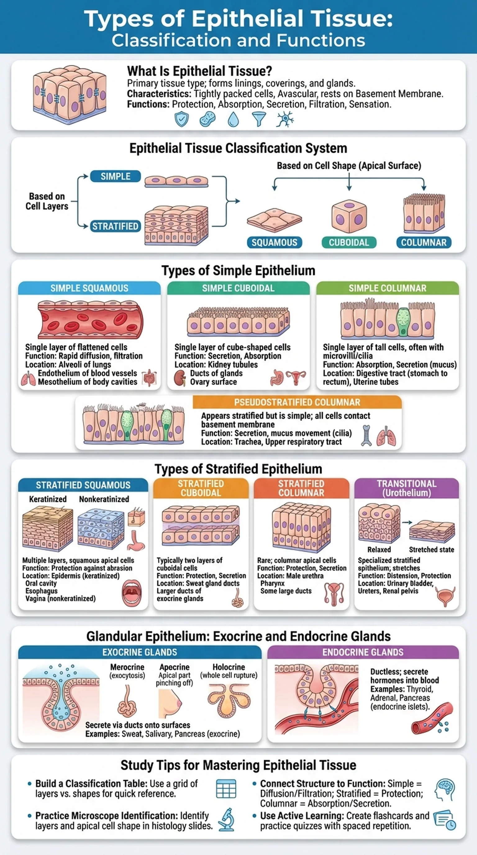
Epithelial tissue is one of the four primary tissue types in the human body, alongside connective, muscle, and nervous tissue. It forms the linings and coverings of virtually every body surface, from the skin that protects us from the external environment to the delicate lining of the small intestine that absorbs nutrients. Understanding epithelial tissue is fundamental for students of anatomy, histology, pathology, and medicine because alterations in epithelial cells are at the root of many diseases, including the vast majority of human cancers (carcinomas).

All types of epithelium share several defining characteristics. First, epithelial cells are tightly packed with minimal extracellular matrix between them, connected by specialized junctions such as tight junctions, adherens junctions, desmosomes, and gap junctions. Second, epithelial tissue is avascular, meaning it lacks its own blood supply and must receive nutrients by diffusion from the underlying connective tissue. Third, all epithelial tissue rests on a basement membrane, a thin extracellular layer composed of a basal lamina and a reticular lamina that anchors the epithelium to the connective tissue below.

Epithelial tissue performs a wide range of functions depending on its location: protection against mechanical abrasion, pathogens, and chemical damage; absorption of nutrients and ions; secretion of mucus, hormones, and enzymes; filtration of blood in the kidneys; and sensation through specialized neuroepithelial cells. The epithelial tissue classification system organizes these diverse tissues based on two criteria: the number of cell layers and the shape of the cells at the free (apical) surface. This dual classification scheme produces the major types of epithelium that every anatomy student must master.

Key Terms

Epithelial Tissue

One of the four primary body tissues, characterized by tightly packed cells that line body surfaces, cavities, and glands, resting on a basement membrane.

Basement Membrane

A thin extracellular layer composed of a basal lamina and reticular lamina that anchors epithelial tissue to the underlying connective tissue.

Tight Junctions

Cell junctions that form a seal between adjacent epithelial cells, preventing the passage of molecules through the intercellular space.

Avascular

Lacking blood vessels; epithelial tissue receives nutrients by diffusion from capillaries in the underlying connective tissue.

Carcinoma

A malignant tumor arising from epithelial tissue, representing the most common category of cancer in humans.

Epithelial Tissue Classification: Layers and Cell Shapes

The epithelial tissue classification system is built on two fundamental criteria: the number of cell layers and the shape of the cells at the apical (free) surface. Mastering this classification is the key to identifying every type of epithelium you will encounter in histology.

Regarding cell layers, epithelial tissues are classified as either simple or stratified. Simple epithelium consists of a single layer of cells, with every cell in direct contact with the basement membrane. This arrangement is ideally suited for functions that require rapid passage of substances, such as absorption, filtration, and secretion. Stratified epithelium consists of two or more layers of cells, with only the deepest layer (the basal layer) touching the basement membrane. The multiple layers provide protection against abrasion and mechanical stress, making stratified epithelium common in areas subjected to wear and tear.

Regarding cell shape, three primary forms are recognized. Squamous cells are flat and scale-like, with a width much greater than their height. Cuboidal cells are roughly cube-shaped, with approximately equal width and height. Columnar cells are tall and column-shaped, with a height significantly greater than their width. The shape of cells at the apical surface determines the descriptive name, even in stratified tissues where the basal cells may differ in shape from the surface cells.

Combining these two criteria produces the major types of epithelium: simple squamous, simple cuboidal, simple columnar, stratified squamous, stratified cuboidal, and stratified columnar. In addition, two special categories exist that do not fit neatly into this grid: pseudostratified columnar epithelium and transitional epithelium (urothelium). This comprehensive epithelial tissue classification system allows histologists and clinicians to identify tissues precisely and correlate their structure with their function.

Key Terms

Epithelial Tissue Classification

The systematic categorization of epithelial tissues based on the number of cell layers (simple vs. stratified) and the shape of apical cells (squamous, cuboidal, columnar).

Squamous

A cell shape that is flat and scale-like, with a width much greater than its height.

Cuboidal

A cell shape that is roughly cube-shaped, with approximately equal width and height.

Columnar

A cell shape that is tall and column-like, with a height significantly greater than its width.

Pseudostratified Epithelium

A type of epithelium that appears to have multiple layers because cell nuclei sit at different heights, but all cells contact the basement membrane.

Types of Simple Epithelium

Simple epithelium, consisting of a single cell layer resting on the basement membrane, is found in locations where thin barriers are needed for diffusion, filtration, absorption, or secretion. The three main types of simple epithelium are simple squamous, simple cuboidal, and simple columnar, each optimized for specific physiological roles.

Simple squamous epithelium is composed of a single layer of flattened cells that form a thin, smooth surface. It lines the air sacs (alveoli) of the lungs, where gas exchange occurs by diffusion across the thinnest possible barrier. It also forms the endothelium lining blood vessels and the mesothelium lining body cavities (pleural, pericardial, and peritoneal). Its extreme thinness makes simple squamous epithelium ideal for passive transport processes but poorly suited for protection.

Simple cuboidal epithelium consists of a single layer of cube-shaped cells and is found in kidney tubules, the surface of the ovary, and the ducts of many glands. Its primary functions include secretion and absorption. In the proximal convoluted tubule of the kidney, simple cuboidal cells have microvilli that increase surface area for reabsorption of water, ions, and nutrients from the glomerular filtrate.

Simple columnar epithelium is a single layer of tall, column-shaped cells that lines most of the gastrointestinal tract, from the stomach to the rectum. In the small intestine, these cells bear dense microvilli forming a brush border that dramatically increases the absorptive surface area. Many simple columnar cells also contain goblet cells, specialized mucus-secreting cells that lubricate and protect the epithelial surface. A variant, pseudostratified columnar epithelium, lines the trachea and upper respiratory tract. Although it appears stratified because nuclei are at different levels, every cell contacts the basement membrane, making it technically a type of simple epithelium with cilia that sweep mucus and trapped particles upward.

Key Terms

Simple Epithelium

Epithelial tissue consisting of a single layer of cells, all in contact with the basement membrane, optimized for diffusion, absorption, filtration, and secretion.

Endothelium

Simple squamous epithelium that lines the interior surface of blood vessels and lymphatic vessels.

Mesothelium

Simple squamous epithelium that lines the pleural, pericardial, and peritoneal body cavities.

Goblet Cells

Specialized mucus-secreting cells interspersed among columnar epithelial cells, particularly abundant in the intestinal and respiratory epithelia.

Microvilli

Tiny finger-like projections on the apical surface of epithelial cells that increase surface area for absorption and secretion.

Types of Stratified Epithelium

Stratified epithelium consists of two or more layers of cells and is found in regions of the body that are subject to abrasion, mechanical stress, or chemical insult. The multiple cell layers provide a protective barrier that can be continuously renewed as surface cells are shed and replaced by cells from the basal layer. The major types of stratified epithelium are stratified squamous, stratified cuboidal, stratified columnar, and transitional epithelium.

Stratified squamous epithelium is by far the most abundant stratified type. It exists in two forms: keratinized and nonkeratinized. Keratinized stratified squamous epithelium forms the epidermis of the skin, where the surface cells are dead, filled with the tough protein keratin, and flattened into a waterproof barrier. This keratinized layer protects underlying tissues from desiccation, pathogens, and physical damage. Nonkeratinized stratified squamous epithelium lines moist surfaces such as the oral cavity, esophagus, and vagina. The surface cells remain nucleated and moist, providing protection against abrasion while allowing flexibility.

Stratified cuboidal epithelium is relatively rare and is found primarily in the ducts of sweat glands and the larger ducts of certain exocrine glands. It typically consists of only two cell layers and provides moderate protection along with a secretory or absorptive function. Stratified columnar epithelium is even rarer, occurring in the male urethra, parts of the pharynx, and some large excretory ducts. Like stratified cuboidal epithelium, it usually has only two or three cell layers.

Transitional epithelium, also called urothelium, is a specialized stratified epithelium found exclusively in the urinary system, lining the renal pelvis, ureters, urinary bladder, and proximal urethra. Its unique feature is the ability to stretch: when the bladder is empty, the cells appear rounded and multilayered, but when the bladder is full, the tissue thins and the cells flatten. This elasticity allows the urinary tract to accommodate varying volumes of urine without rupturing, demonstrating how epithelial tissue classification reflects functional specialization.

Key Terms

Stratified Epithelium

Epithelial tissue consisting of two or more cell layers, with only the basal layer contacting the basement membrane, providing protection against abrasion and stress.

Keratinized Epithelium

Stratified squamous epithelium whose surface cells are dead and filled with keratin, forming a tough, waterproof barrier as seen in the epidermis.

Transitional Epithelium (Urothelium)

A specialized stratified epithelium of the urinary tract that can stretch and change shape to accommodate volume changes.

Keratin

A tough, fibrous structural protein that fills the surface cells of keratinized stratified squamous epithelium, providing waterproofing and mechanical protection.

Glandular Epithelium: Exocrine and Endocrine Glands

Glandular epithelium is a specialized type of epithelial tissue composed of cells that are adapted for secretion. During embryological development, glandular epithelium arises from surface epithelium that invaginates into the underlying connective tissue. Glands are classified as either exocrine or endocrine based on whether they retain a connection to the surface epithelium.

Exocrine glands maintain a duct that delivers their secretory products to an epithelial surface. Examples include sweat glands, salivary glands, the pancreas (exocrine portion), and mammary glands. Exocrine glands are further classified by their duct structure (simple vs. compound) and by the shape of their secretory units (tubular, acinar, or tubuloacinar). They are also categorized by their mode of secretion: merocrine glands (such as eccrine sweat glands) secrete products by exocytosis without losing cellular material; apocrine glands release secretions along with a portion of the apical cytoplasm; and holocrine glands (such as sebaceous glands) release their entire cell contents as the cell disintegrates.

Endocrine glands lose their ductal connection during development and secrete hormones directly into the bloodstream. Examples include the thyroid gland, adrenal glands, and the islets of Langerhans in the pancreas. The pancreas is a unique organ possessing both exocrine and endocrine functions, making it a mixed gland. Understanding glandular epithelium is essential because many types of epithelium have secretory functions, and tumors arising from glandular epithelial tissue (adenocarcinomas) are among the most common cancers. Epithelial tissue in glandular form illustrates the remarkable diversity of this tissue type, extending its role far beyond simple surface coverage to include complex regulatory and secretory functions throughout the body.

Key Terms

Exocrine Glands

Glands that secrete their products onto an epithelial surface via ducts, such as sweat glands, salivary glands, and the exocrine pancreas.

Endocrine Glands

Ductless glands that secrete hormones directly into the bloodstream, such as the thyroid and adrenal glands.

Merocrine Secretion

A mode of secretion in which products are released by exocytosis without loss of cellular material, as in eccrine sweat glands.

Holocrine Secretion

A mode of secretion in which the entire cell disintegrates to release its contents, as in sebaceous glands.

Adenocarcinoma

A malignant tumor arising from glandular epithelial tissue, representing a common subtype of carcinoma.

Study Tips for Mastering Types of Epithelial Tissue

Mastering the types of epithelial tissue requires a systematic approach that leverages the logical classification framework built into the topic. Because epithelial tissue classification follows clear rules based on cell layers and cell shape, students can use pattern recognition rather than brute-force memorization. Here are effective strategies for learning and retaining this material.

First, build a classification table. Create a grid with cell layers (simple vs. stratified) on one axis and cell shapes (squamous, cuboidal, columnar) on the other. Fill in each cell with the tissue name, a representative location, and its primary function. Add rows for the special categories: pseudostratified columnar and transitional epithelium. This single table provides a comprehensive overview of all types of epithelium and serves as an excellent quick-reference study tool.

Second, learn to identify each type of epithelium under the microscope. Histology exams will present you with tissue slides, and you must classify the epithelium by systematically asking two questions: How many layers are there (simple or stratified)? What is the shape of the apical cells (squamous, cuboidal, or columnar)? Practicing with labeled histology images trains your eye to distinguish between simple epithelium and stratified epithelium at a glance. Pay attention to nuclear shape as a clue: flat nuclei suggest squamous cells, round nuclei suggest cuboidal cells, and elongated nuclei suggest columnar cells.

Third, connect structure to function for each type. Simple squamous equals thin barrier for diffusion. Stratified squamous equals protection against abrasion. Simple columnar equals absorption and secretion. These structure-function pairings are the most commonly tested concepts. Finally, use active learning tools such as LectureScribe to generate flashcards and practice quizzes from your lecture notes on epithelial tissue. Spaced repetition of the classification system will build permanent recall that translates to confident performance on anatomy and histology examinations.

Key Terms

Types of Epithelium

The different categories of epithelial tissue classified by cell layers (simple, stratified) and cell shape (squamous, cuboidal, columnar), plus special types.

Active Recall

A study technique where learners actively retrieve information from memory rather than passively reviewing notes.

Spaced Repetition

A learning strategy that involves reviewing material at increasing intervals to strengthen long-term memory retention.

Frequently Asked Questions

What is epithelial tissue and what does it do?

Epithelial tissue is one of the four primary body tissues, consisting of tightly packed cells that line body surfaces, cavities, and organs. It performs functions including protection, absorption, secretion, filtration, and sensation. All epithelial tissue rests on a basement membrane and is avascular.

What are the main types of epithelium?

The main types of epithelium are simple squamous, simple cuboidal, simple columnar, stratified squamous, stratified cuboidal, stratified columnar, pseudostratified columnar, and transitional epithelium. They are classified by the number of cell layers and the shape of the apical cells.

How does epithelial tissue classification work?

Epithelial tissue classification is based on two criteria: the number of cell layers (simple for one layer, stratified for multiple layers) and the shape of the cells at the apical surface (squamous for flat, cuboidal for cube-shaped, columnar for tall). Combining these criteria produces the major tissue types.

What is the difference between simple epithelium and stratified epithelium?

Simple epithelium consists of a single layer of cells, all touching the basement membrane, and is suited for absorption, filtration, and secretion. Stratified epithelium has two or more cell layers, with only the basal layer touching the basement membrane, and is designed primarily for protection against abrasion.

Where is stratified squamous epithelium found in the body?

Stratified squamous epithelium is found in areas subject to abrasion. The keratinized form makes up the epidermis of the skin. The nonkeratinized form lines the oral cavity, esophagus, vagina, and anal canal. It is the most widespread type of stratified epithelium in the body.

What is pseudostratified columnar epithelium?

Pseudostratified columnar epithelium appears to have multiple layers because its cell nuclei lie at different heights, but all cells contact the basement membrane, making it technically simple. It lines the trachea and upper respiratory tract, where its cilia move mucus and trapped particles.

Why is epithelial tissue important in cancer biology?

Epithelial tissue gives rise to carcinomas, which account for approximately 80-90% of all human cancers. Because epithelial cells line surfaces exposed to environmental insults and divide frequently to replace shed cells, they are particularly susceptible to the mutations that drive malignant transformation.

Study Anatomy Smarter

Upload your lecture notes and get AI-generated flashcards, quizzes, infographics, and study guides tailored to your curriculum.

Try LectureScribe Free

Create Study Materials

Transform your Anatomy lectures into flashcards, quizzes, and visual study guides with AI.

  • AI-generated flashcards & quizzes
  • Visual infographics from notes
  • Interactive study shorts
Start Studying Free

Quick Facts

SubjectAnatomy
Word Count2,500+
Slides5
InfographicIncluded
AuthorLectureScribe

Master Anatomy with LectureScribe

Upload your lecture notes and get AI-powered flashcards, quizzes, infographics, and study guides in minutes.