LectureScribe Logo
lecturescribe.io

LOCK IN VALENTINE PRICING BEFORE SALE ENDS!

Use code — New joiners get 40% OFF on annual plans

Plans will have higher prices after this sale ends

0days
:
0hours
:
0minutes
:
0seconds
Anatomy2,500+ words5 slides
🦴

Types of Joints in the Body: Synovial, Fibrous & Cartilaginous

Learn the types of joints in the body including synovial joints, fibrous joints, and cartilaginous joints. Understand joint classification by structure and function.

L
LectureScribeAI-Powered Study Platform

Study Infographic

Complete types of joints overview illustrating structural classification, functional categories, and clinical examples of fibrous, cartilaginous, and synovial joints.

Download
Joint classification diagram showing fibrous, cartilaginous, and synovial joints with structural features and examples

Interactive Study Short

Swipe through 5 slides about Types of Joints in the Body: Synovial, Fibrous & Cartilaginous

1 / 5

Introduction to Joint Classification

Joints, also known as articulations, are the points where two or more bones meet in the body. Understanding the types of joints is fundamental to the study of human anatomy because joints determine the range and type of movement possible at each skeletal connection. Joint classification provides a systematic framework for organizing the hundreds of articulations in the human body according to their structural characteristics and functional capabilities. Without joints, the rigid skeleton would be incapable of the fluid, coordinated movements that define human locomotion, manual dexterity, and postural control.

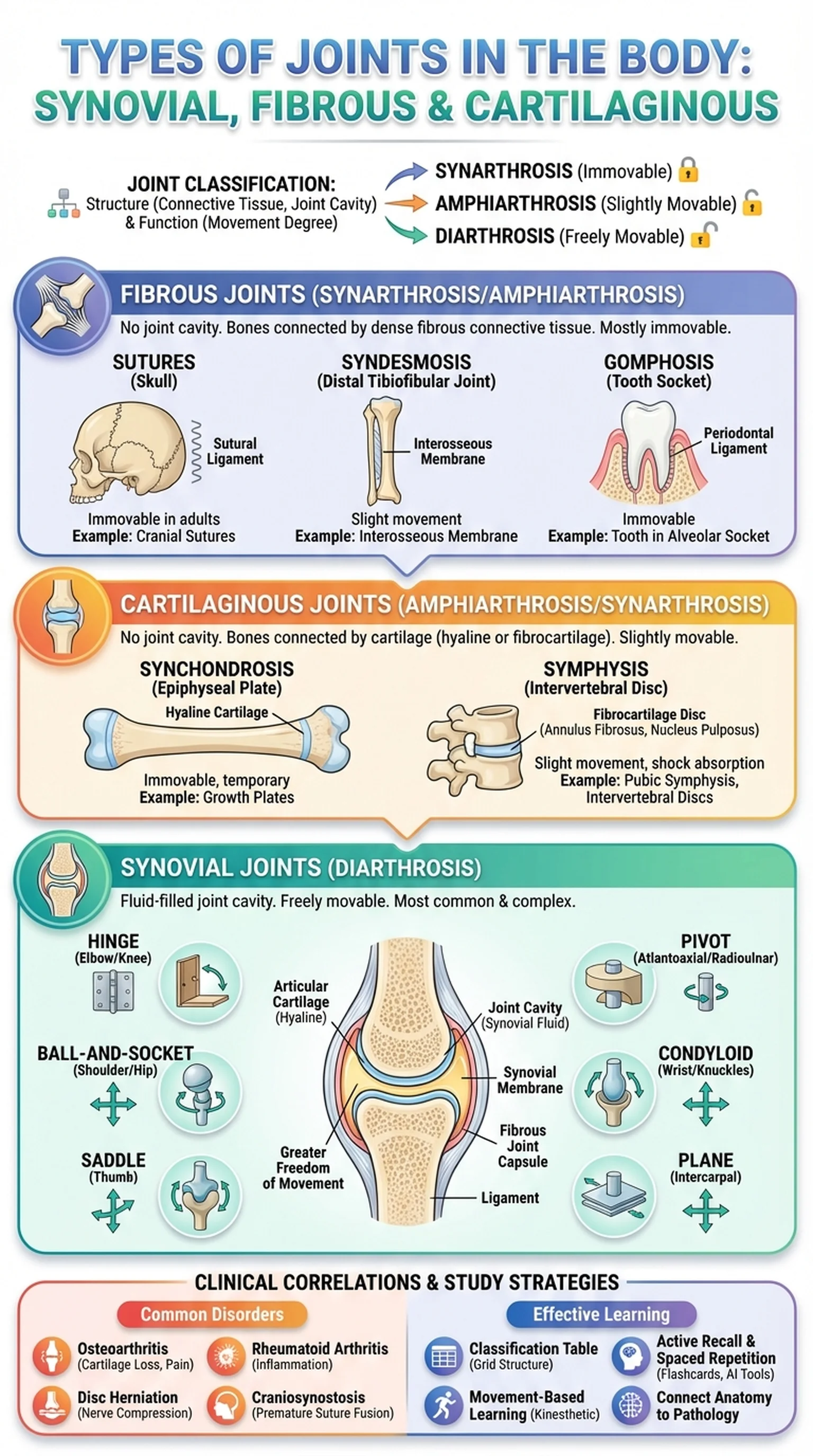
Joint classification can be approached from two complementary perspectives: structural and functional. Structural classification groups joints by the type of connective tissue that binds the bones together and whether a joint cavity is present. The three structural categories are fibrous joints (connected by dense fibrous tissue, no joint cavity), cartilaginous joints (connected by cartilage, no joint cavity), and synovial joints (separated by a fluid-filled joint cavity lined with synovial membrane). Functional classification groups joints by their degree of movement: synarthroses are immovable joints, amphiarthroses are slightly movable joints, and diarthroses are freely movable joints.

These two classification systems overlap in predictable ways. Most fibrous joints are synarthroses, most cartilaginous joints are amphiarthroses, and all synovial joints are diarthroses. Learning both systems simultaneously provides a more complete understanding of the types of joints and their clinical relevance. For students preparing for anatomy exams and board certifications, joint classification is a recurring theme that integrates concepts from musculoskeletal anatomy, kinesiology, and orthopedic medicine.

Key Terms

Types of Joints

The classification of skeletal articulations based on structural composition (fibrous, cartilaginous, synovial) and functional mobility (synarthrosis, amphiarthrosis, diarthrosis).

Joint Classification

The systematic categorization of joints by structure (type of connective tissue and presence of a joint cavity) and function (degree of permitted movement).

Articulation

The point of contact between two or more bones, also known as a joint, which may allow varying degrees of movement.

Synarthrosis

An immovable joint, such as the sutures of the skull, where bones are tightly bound and no movement occurs.

Fibrous Joints: Structure, Types, and Examples

Fibrous joints are articulations in which the bones are connected by dense fibrous connective tissue, with no joint cavity present. Because the bones are tightly bound by collagen fibers, fibrous joints generally permit little to no movement, making most of them functionally classified as synarthroses (immovable) or amphiarthroses (slightly movable). Fibrous joints are found primarily in locations where stability and protection are more important than mobility.

There are three subtypes of fibrous joints: sutures, syndesmoses, and gomphoses. Sutures are the interlocking fibrous joints found exclusively between the flat bones of the skull. The serrated, interdigitating edges of adjacent skull bones are connected by a thin layer of dense fibrous tissue called the sutural ligament. Sutures are functionally immovable (synarthroses) in adults, though they allow slight flexibility during infancy to accommodate brain growth and passage through the birth canal. Over time, many sutures ossify completely to form synostoses, where the fibrous tissue is replaced by bone.

Syndesmoses are fibrous joints in which the bones are connected by a ligament or an interosseous membrane spanning a greater distance than in sutures. The distal tibiofibular joint is a classic example, where the interosseous membrane connects the tibia and fibula, permitting slight rotational movement (making it an amphiarthrosis). The interosseous membrane of the forearm between the radius and ulna is another important syndesmosis. Gomphoses are specialized fibrous joints found only between the roots of teeth and the alveolar sockets of the mandible and maxilla. The periodontal ligament anchors each tooth in its socket, creating a joint that is functionally immovable under normal conditions. Recognizing fibrous joints and their subtypes is essential for a complete understanding of joint classification and the diverse types of joints in the body.

Key Terms

Fibrous Joints

Joints in which bones are connected by dense fibrous connective tissue without a joint cavity, typically permitting little or no movement.

Sutures

Fibrous joints found between skull bones, characterized by interlocking edges connected by sutural ligaments, functionally immovable in adults.

Syndesmosis

A fibrous joint in which bones are connected by a ligament or interosseous membrane, allowing slight movement, as in the distal tibiofibular joint.

Gomphosis

A specialized fibrous joint between a tooth root and its alveolar socket, anchored by the periodontal ligament.

Cartilaginous Joints: Structure, Types, and Examples

Cartilaginous joints are articulations in which bones are connected by either hyaline cartilage or fibrocartilage, with no joint cavity present. These joints typically allow limited movement, placing most of them in the amphiarthrosis (slightly movable) functional category, though some are synarthroses. Cartilaginous joints are found at locations in the body where moderate stability is needed alongside the capacity for slight deformation or compression.

There are two subtypes of cartilaginous joints: synchondroses and symphyses. Synchondroses are joints where the connecting tissue is hyaline cartilage. The most important examples are the epiphyseal plates (growth plates) in growing long bones, where a band of hyaline cartilage separates the epiphysis from the diaphysis, allowing longitudinal bone growth. Synchondroses are functionally immovable (synarthroses) and are temporary in many cases, as the hyaline cartilage is eventually replaced by bone when growth is complete (forming a synostosis). The first sternocostal joint, where the first rib articulates with the manubrium of the sternum via hyaline cartilage, is a permanent synchondrosis.

Symphyses are cartilaginous joints in which the bones are connected by a disc or pad of fibrocartilage, often with a thin layer of hyaline cartilage covering the articular surfaces of the adjacent bones. Symphyses are slightly movable (amphiarthroses) and are designed to absorb compressive forces and provide resilient cushioning. The pubic symphysis, connecting the two pubic bones at the anterior pelvis, and the intervertebral discs between adjacent vertebral bodies are the most clinically significant examples. The intervertebral discs, with their tough outer annulus fibrosus and gel-like inner nucleus pulposus, permit slight flexion, extension, and rotation at each spinal level. When a disc herniates, the nucleus pulposus protrudes through the annulus, potentially compressing spinal nerves. Mastering cartilaginous joints is vital for understanding both normal joint classification and the pathology of common musculoskeletal conditions.

Key Terms

Cartilaginous Joints

Joints in which bones are connected by hyaline cartilage (synchondroses) or fibrocartilage (symphyses), typically allowing limited or no movement.

Synchondrosis

A cartilaginous joint connected by hyaline cartilage, such as the epiphyseal plate, that is functionally immovable and often temporary.

Symphysis

A cartilaginous joint connected by a fibrocartilage disc, such as the pubic symphysis or intervertebral disc, that allows slight movement.

Intervertebral Disc

A fibrocartilaginous pad between vertebral bodies that absorbs compressive forces and permits limited spinal movement.

Synovial Joints: The Freely Movable Joints

Synovial joints are the most common and most complex types of joints in the human body. They are the only joints that possess a fluid-filled joint cavity, and they are all functionally classified as diarthroses (freely movable). Synovial joints are found at virtually every location where significant movement is required, including the shoulder, hip, knee, elbow, wrist, and fingers. Their structural sophistication allows for an extraordinary range of motion while maintaining stability and minimizing friction.

The defining features of synovial joints include articular cartilage, a joint (synovial) cavity, a synovial membrane, synovial fluid, and a fibrous joint capsule. Articular cartilage, composed of hyaline cartilage, covers the ends of the articulating bones, providing a smooth, low-friction surface that absorbs shock. The synovial membrane lines the inner surface of the joint capsule and secretes synovial fluid, a viscous liquid that lubricates the joint surfaces, nourishes the avascular articular cartilage, and removes metabolic waste. The fibrous joint capsule, reinforced by ligaments, encloses the joint cavity and provides structural stability. Many synovial joints also contain accessory structures such as menisci, bursae, fat pads, and tendons that enhance joint function.

Synovial joints are further classified into six subtypes based on the shape of their articular surfaces and the types of movement they permit. Plane (gliding) joints allow sliding movements (intercarpal joints). Hinge joints permit flexion and extension in one plane (elbow, knee). Pivot joints allow rotation around a single axis (atlantoaxial joint, proximal radioulnar joint). Condyloid (ellipsoid) joints permit flexion, extension, abduction, and adduction but not rotation (metacarpophalangeal joints). Saddle joints allow the same movements as condyloid joints plus greater freedom (first carpometacarpal joint of the thumb). Ball-and-socket joints are the most mobile, permitting movement in all planes including rotation (shoulder, hip). This subtype classification of synovial joints is essential for understanding joint classification and predicting the movements possible at any given articulation.

Key Terms

Synovial Joints

Freely movable joints characterized by a fluid-filled joint cavity, articular cartilage, a synovial membrane, and a fibrous capsule, found throughout the body.

Synovial Fluid

A viscous liquid secreted by the synovial membrane that lubricates articular surfaces, nourishes cartilage, and reduces friction within synovial joints.

Ball-and-Socket Joint

A synovial joint subtype allowing multiaxial movement including rotation, exemplified by the shoulder and hip joints.

Hinge Joint

A synovial joint subtype permitting uniaxial flexion and extension, exemplified by the elbow and knee joints.

Articular Cartilage

A layer of hyaline cartilage covering the bone surfaces within a synovial joint, providing a smooth, low-friction surface for movement.

Clinical Correlations: Joint Disorders and Injuries

Understanding the types of joints and their structural features is directly relevant to clinical medicine, as joint disorders and injuries are among the most common reasons patients seek medical care. The clinical significance of joint classification becomes apparent when considering how specific structural features of fibrous joints, cartilaginous joints, and synovial joints predispose them to characteristic pathologies.

Synovial joints, being the most mobile and structurally complex, are the most susceptible to injury and degenerative disease. Osteoarthritis, the most common form of arthritis, involves progressive degradation of articular cartilage within synovial joints, leading to pain, stiffness, and reduced range of motion. It most frequently affects weight-bearing joints such as the knee and hip. Rheumatoid arthritis is an autoimmune condition in which the immune system attacks the synovial membrane, causing inflammation, joint destruction, and deformity. Ligament tears, meniscal injuries, and dislocations are traumatic injuries specific to synovial joints and are particularly common in athletes.

Cartilaginous joints also have characteristic pathologies. Herniation of intervertebral discs (symphyses) is one of the most prevalent musculoskeletal conditions, often causing radiculopathy when the displaced material compresses a spinal nerve root. The pubic symphysis can become dysfunctional during pregnancy due to hormonal-mediated ligament laxity. Fibrous joints are generally resistant to injury because of their immobility, but premature fusion of cranial sutures (craniosynostosis) in infants can restrict brain growth and require surgical intervention. A comprehensive understanding of the types of joints, from fibrous joints to cartilaginous joints to synovial joints, enables clinicians to predict injury patterns, interpret imaging studies, and develop targeted treatment plans for joint pathology.

Key Terms

Osteoarthritis

A degenerative joint disease characterized by progressive loss of articular cartilage in synovial joints, leading to pain and functional impairment.

Rheumatoid Arthritis

An autoimmune disorder that targets the synovial membrane of joints, causing chronic inflammation, cartilage destruction, and joint deformity.

Disc Herniation

A condition in which the nucleus pulposus of an intervertebral disc (a symphysis-type cartilaginous joint) protrudes through the annulus fibrosus, potentially compressing spinal nerves.

Craniosynostosis

Premature fusion of one or more cranial sutures (fibrous joints) in an infant, potentially restricting brain growth and requiring surgical correction.

Study Strategies for Joint Anatomy

The types of joints are a core topic on anatomy examinations and medical licensing tests including the USMLE Step 1, COMLEX, and MCAT. Questions often require students to classify joints by structure and function, identify specific joint types from clinical or histological descriptions, and apply joint anatomy to clinical scenarios. Here are effective strategies for mastering this material.

First, build a classification table. Create a grid with three columns for fibrous joints, cartilaginous joints, and synovial joints. For each category, list the subtypes, defining structural features, functional classification (synarthrosis, amphiarthrosis, or diarthrosis), and two to three clinical examples. This single table captures the essential framework of joint classification and serves as a rapid review tool before exams. Having this organizational structure in mind makes it easy to categorize any joint you encounter.

Second, use the six subtypes of synovial joints as a movement-based learning tool. For each subtype (plane, hinge, pivot, condyloid, saddle, ball-and-socket), identify the types of movement permitted and practice locating examples on your own body. Move the joint through its range of motion while naming the movements (flexion, extension, abduction, adduction, rotation, circumduction). This kinesthetic approach engages multiple learning modalities and makes the abstract concept of joint classification tangible and memorable.

Third, connect each joint type to its most important clinical condition. Synovial joints link to osteoarthritis and ligament injuries; cartilaginous joints link to disc herniation; fibrous joints link to craniosynostosis. This pairing of anatomy with pathology mirrors how board exams test the material and reinforces the clinical relevance of the types of joints. Finally, use AI-powered study platforms like LectureScribe to generate flashcards, practice questions, and slide decks from your notes on joint classification, synovial joints, fibrous joints, and cartilaginous joints. Active recall and spaced repetition are the most effective methods for retaining the detailed anatomical knowledge required for exam success.

Key Terms

Active Recall

A study technique involving deliberate retrieval of information from memory, proven to be more effective than passive review for long-term retention.

Spaced Repetition

A learning strategy that spaces review sessions at increasing intervals to optimize memory consolidation over time.

USMLE Step 1

A standardized medical licensing exam testing foundational science knowledge, including musculoskeletal anatomy and joint classification.

Frequently Asked Questions

What are the three main types of joints in the body?

The three main types of joints are fibrous joints (connected by dense fibrous tissue), cartilaginous joints (connected by cartilage), and synovial joints (featuring a fluid-filled joint cavity). This structural classification is the foundation of joint anatomy.

What are synovial joints and why are they important?

Synovial joints are freely movable joints characterized by a joint cavity filled with synovial fluid, articular cartilage, and a fibrous capsule. They are the most common joint type and are found at the shoulder, hip, knee, and elbow, enabling the wide range of movements needed for daily activities.

What are fibrous joints and where are they found?

Fibrous joints are articulations where bones are connected by dense fibrous tissue without a joint cavity. They include sutures (skull), syndesmoses (distal tibiofibular joint), and gomphoses (teeth in their sockets). Most fibrous joints are immovable.

What are cartilaginous joints?

Cartilaginous joints are joints connected by cartilage without a joint cavity. Synchondroses are connected by hyaline cartilage (epiphyseal plates), while symphyses are connected by fibrocartilage (intervertebral discs, pubic symphysis). They allow limited or no movement.

How is joint classification organized?

Joint classification uses two complementary systems. Structural classification groups joints by connective tissue type (fibrous, cartilaginous, synovial). Functional classification groups joints by degree of movement (synarthrosis for immovable, amphiarthrosis for slightly movable, diarthrosis for freely movable).

What are the six subtypes of synovial joints?

The six subtypes are plane (gliding), hinge (flexion/extension), pivot (rotation), condyloid (biaxial), saddle (biaxial with greater freedom), and ball-and-socket (multiaxial). Each subtype is defined by the shape of the articular surfaces and the movements permitted.

What common diseases affect joints?

Common joint diseases include osteoarthritis (cartilage degeneration in synovial joints), rheumatoid arthritis (autoimmune synovial inflammation), disc herniation (cartilaginous joint pathology), and craniosynostosis (premature fusion of fibrous skull sutures).

Study Anatomy Smarter

Upload your lecture notes and get AI-generated flashcards, quizzes, infographics, and study guides tailored to your curriculum.

Try LectureScribe Free

Create Study Materials

Transform your Anatomy lectures into flashcards, quizzes, and visual study guides with AI.

  • AI-generated flashcards & quizzes
  • Visual infographics from notes
  • Interactive study shorts
Start Studying Free

Quick Facts

SubjectAnatomy
Word Count2,500+
Slides5
InfographicIncluded
AuthorLectureScribe

Master Anatomy with LectureScribe

Upload your lecture notes and get AI-powered flashcards, quizzes, infographics, and study guides in minutes.