LectureScribe Logo
lecturescribe.io

LOCK IN VALENTINE PRICING BEFORE SALE ENDS!

Use code — New joiners get 40% OFF on annual plans

Plans will have higher prices after this sale ends

0days
:
0hours
:
0minutes
:
0seconds
Neuroscience2,500+ words5 slides
🧠

Neuroanatomy: Brain Regions, Pathways & Clinical Correlations

Master neuroanatomy including brain regions, neural pathways, basal ganglia, thalamus, and cerebellum. Comprehensive guide with clinical correlations for medical and neuroscience students.

L
LectureScribeAI-Powered Study Platform

Study Infographic

Complete neuroanatomy reference infographic illustrating brain regions, key neural pathways, and clinical correlations for the thalamus, basal ganglia, and cerebellum.

Download
Neuroanatomy diagram showing major brain regions including cerebral cortex, thalamus, basal ganglia, cerebellum, and their neural pathways with clinical correlations

Interactive Study Short

Swipe through 5 slides about Neuroanatomy: Brain Regions, Pathways & Clinical Correlations

1 / 5

Introduction to Neuroanatomy and Brain Organization

Neuroanatomy is the branch of anatomy dedicated to the study of the structure and organization of the nervous system. For medical students, neuroscience majors, and allied health professionals, a solid understanding of neuroanatomy provides the essential framework for interpreting clinical findings, localizing lesions, and understanding the neural basis of behavior. The human brain contains approximately 86 billion neurons organized into distinct brain regions, each with specialized functions and interconnected by elaborate neural pathways.

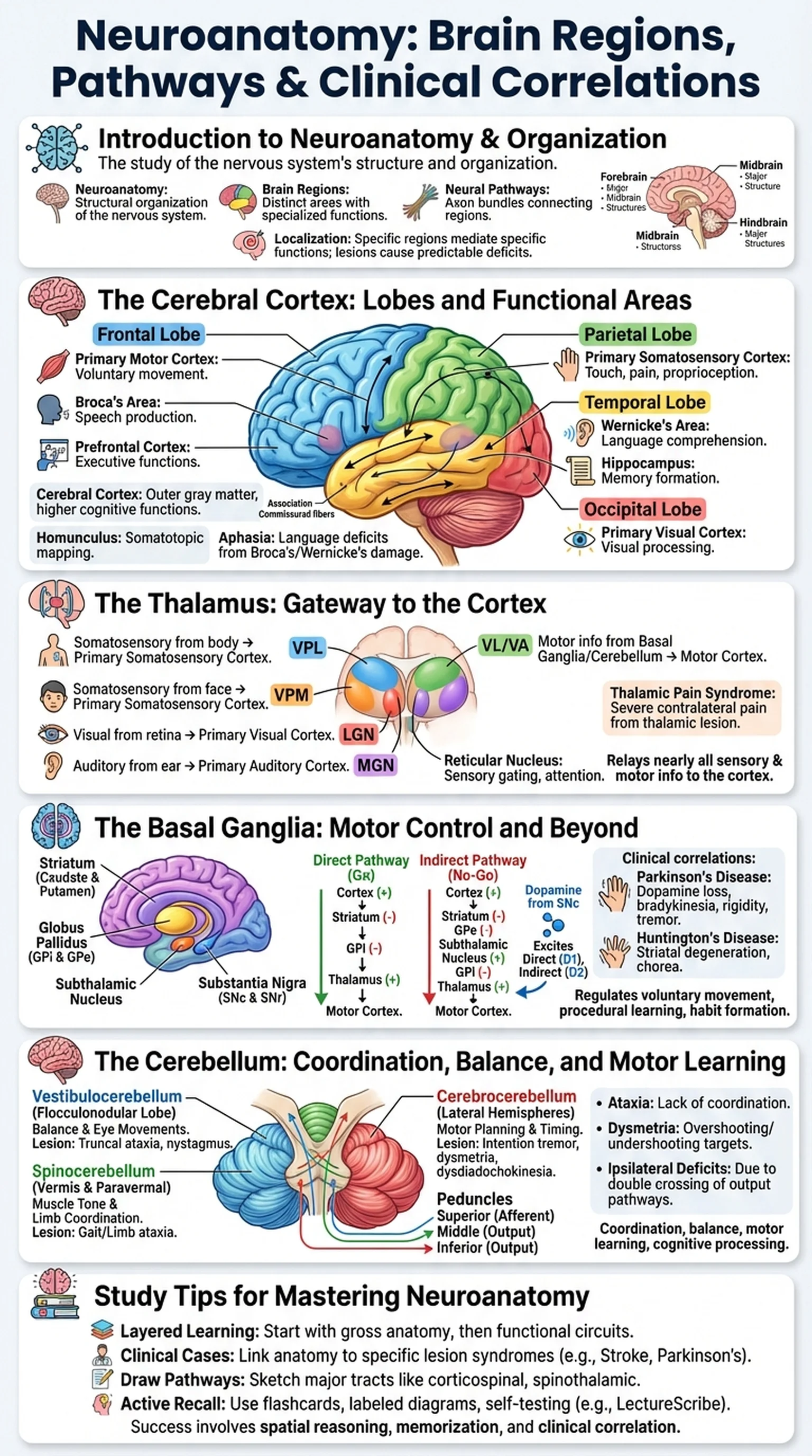
The brain can be divided into several major structures based on embryological development. The forebrain (prosencephalon) gives rise to the cerebral cortex, thalamus, hypothalamus, and basal ganglia. The midbrain (mesencephalon) contains structures involved in visual and auditory reflexes, motor control, and arousal. The hindbrain (rhombencephalon) comprises the pons, medulla oblongata, and cerebellum. Each of these brain regions contributes uniquely to the integrated functioning of the nervous system.

Understanding neuroanatomy requires learning both structure and connectivity. Individual brain regions do not operate in isolation; instead, they communicate through bundles of axons called neural pathways or tracts. These pathways can be ascending (carrying sensory information to the cortex), descending (carrying motor commands from the cortex to the spinal cord), or associative (connecting different cortical areas within the same hemisphere). The clinical relevance of neuroanatomy lies in the principle of localization: specific lesions in specific brain regions produce predictable clinical deficits. This principle allows clinicians to pinpoint the location of strokes, tumors, and degenerative diseases based on a patient's neurological examination.

Key Terms

Neuroanatomy

The study of the structural organization of the nervous system, including the brain, spinal cord, and peripheral nerves.

Brain Regions

Anatomically and functionally distinct areas of the brain, including the cerebral cortex, thalamus, basal ganglia, cerebellum, brainstem, and others.

Neural Pathways

Bundles of axons connecting different brain regions, transmitting sensory, motor, and associative information throughout the nervous system.

Localization

The neurological principle that specific brain regions mediate specific functions, allowing clinicians to identify lesion sites from clinical symptoms.

The Cerebral Cortex: Lobes and Functional Areas

The cerebral cortex is the outermost layer of the brain, a sheet of gray matter approximately 2 to 4 millimeters thick that is extensively folded into gyri (ridges) and sulci (grooves). It is divided into four lobes on each hemisphere: the frontal lobe, parietal lobe, temporal lobe, and occipital lobe. Each lobe contains multiple functional areas that serve as key brain regions for specific cognitive, sensory, and motor processes.

The frontal lobe, the largest of the four, extends from the anterior pole to the central sulcus. It contains the primary motor cortex (precentral gyrus), which controls voluntary movement of the contralateral body, organized somatotopically as depicted by the motor homunculus. Anterior to the motor cortex lies the prefrontal cortex, responsible for executive functions including planning, decision-making, working memory, and personality. Broca's area, located in the inferior frontal gyrus of the dominant hemisphere, is essential for speech production. Damage produces expressive (Broca's) aphasia.

The parietal lobe lies posterior to the central sulcus and contains the primary somatosensory cortex (postcentral gyrus), processing touch, proprioception, and pain from the contralateral body. The posterior parietal cortex integrates sensory information for spatial awareness and attention. The temporal lobe, situated laterally below the Sylvian fissure, houses the primary auditory cortex, Wernicke's area for language comprehension, and the hippocampus for memory formation. The occipital lobe, at the posterior pole, contains the primary visual cortex (V1), which receives input from the lateral geniculate nucleus of the thalamus via the optic radiations. These cortical brain regions are interconnected by association fibers, commissural fibers (such as the corpus callosum), and projection fibers that form the complex neural pathways underlying all higher cognitive functions.

Key Terms

Cerebral Cortex

The outer layer of gray matter covering the cerebral hemispheres, responsible for higher cognitive functions, sensory processing, and voluntary motor control.

Primary Motor Cortex

The precentral gyrus of the frontal lobe that initiates voluntary movement of the contralateral body via the corticospinal tract.

Broca's Area

A region in the inferior frontal gyrus of the dominant hemisphere essential for speech production; damage causes expressive aphasia.

Wernicke's Area

A region in the superior temporal gyrus of the dominant hemisphere critical for language comprehension; damage causes receptive aphasia.

Hippocampus

A medial temporal lobe structure essential for the formation of new declarative memories, with extensive connections to cortical brain regions.

The Thalamus: Gateway to the Cortex

The thalamus is a bilateral, egg-shaped structure located in the center of the brain, forming the largest component of the diencephalon. Often described as the gateway or relay station of the brain, the thalamus processes and transmits nearly all sensory and motor information destined for the cerebral cortex. With the exception of olfaction, which bypasses the thalamus and projects directly to cortical areas, every sensory modality synapses in a specific thalamic nucleus before reaching its corresponding cortical area. This makes the thalamus one of the most critical brain regions for conscious sensory experience.

The thalamus is composed of numerous nuclei, each with specific connections and functions. The ventral posterolateral (VPL) nucleus receives somatosensory information from the body via the medial lemniscus and spinothalamic tracts and relays it to the primary somatosensory cortex. The ventral posteromedial (VPM) nucleus relays somatosensory information from the face via the trigeminal pathway. The lateral geniculate nucleus (LGN) processes visual information from the retina and projects to the primary visual cortex. The medial geniculate nucleus (MGN) relays auditory information to the primary auditory cortex. The ventral lateral (VL) and ventral anterior (VA) nuclei receive input from the basal ganglia and cerebellum and relay motor planning information to the motor cortex.

Beyond its relay function, the thalamus plays active roles in attention, consciousness, and sleep-wake regulation. The reticular nucleus of the thalamus acts as a gating mechanism, modulating which sensory information reaches cortical awareness. The intralaminar nuclei contribute to arousal and are part of the ascending reticular activating system. Thalamic lesions can produce devastating clinical syndromes: a stroke affecting the thalamus may cause contralateral sensory loss, thalamic pain syndrome, or even disturbances of consciousness. Understanding the thalamus and its neural pathways to cortical brain regions is therefore essential for clinical neuroanatomy.

Key Terms

Thalamus

A diencephalic structure that relays and processes nearly all sensory and motor information destined for the cerebral cortex, serving as the brain's central relay station.

Lateral Geniculate Nucleus

A thalamic nucleus that receives visual input from the retina and relays it to the primary visual cortex via the optic radiations.

VPL Nucleus

The ventral posterolateral nucleus of the thalamus, relaying somatosensory information from the body to the primary somatosensory cortex.

Reticular Nucleus

A shell-like thalamic nucleus that modulates thalamocortical relay activity, functioning as an attentional gate for sensory information.

Thalamic Pain Syndrome

A clinical condition following thalamic stroke characterized by severe, often burning contralateral pain due to disruption of somatosensory relay circuits.

The Basal Ganglia: Motor Control and Beyond

The basal ganglia are a group of subcortical nuclei situated deep within the cerebral hemispheres that play a pivotal role in the regulation of voluntary movement, procedural learning, habit formation, and reward-based decision-making. The principal components of the basal ganglia include the caudate nucleus, putamen, globus pallidus (internal and external segments), subthalamic nucleus, and substantia nigra (pars compacta and pars reticulata). The caudate and putamen together form the striatum, which serves as the primary input structure of the basal ganglia.

The basal ganglia operate through two major neural pathways that exert opposing effects on motor output. The direct pathway facilitates movement: cortical excitation of the striatum leads to inhibition of the globus pallidus internal segment (GPi), which in turn disinhibits the thalamus, allowing it to excite the motor cortex. The indirect pathway suppresses unwanted movement: cortical activation of the striatum inhibits the globus pallidus external segment (GPe), which disinhibits the subthalamic nucleus, ultimately increasing GPi inhibition of the thalamus. The balance between these two neural pathways determines whether a planned movement is executed or suppressed.

Dopamine from the substantia nigra pars compacta modulates this balance by exciting the direct pathway (via D1 receptors) and inhibiting the indirect pathway (via D2 receptors). Loss of dopaminergic neurons in the substantia nigra disrupts this balance, producing the cardinal symptoms of Parkinson's disease: bradykinesia, rigidity, resting tremor, and postural instability. Conversely, degeneration of striatal neurons, as occurs in Huntington's disease, disproportionately affects the indirect pathway, leading to excessive involuntary movements (chorea). Understanding basal ganglia circuitry and its neural pathways is essential for localizing lesions and comprehending the pharmacological treatment of movement disorders in clinical neuroanatomy.

Key Terms

Basal Ganglia

A group of subcortical nuclei including the striatum, globus pallidus, subthalamic nucleus, and substantia nigra that regulate voluntary movement and procedural learning.

Striatum

The primary input structure of the basal ganglia, comprising the caudate nucleus and putamen, which receives cortical and dopaminergic projections.

Direct Pathway

A basal ganglia circuit that facilitates movement by inhibiting the GPi and disinhibiting the thalamus, promoted by dopamine acting on D1 receptors.

Indirect Pathway

A basal ganglia circuit that suppresses unwanted movement by increasing GPi activity and inhibiting the thalamus, modulated by dopamine at D2 receptors.

Substantia Nigra

A midbrain structure whose pars compacta provides dopaminergic input to the basal ganglia; its degeneration causes Parkinson's disease.

The Cerebellum: Coordination, Balance, and Motor Learning

The cerebellum is a distinctive brain region located in the posterior cranial fossa, dorsal to the brainstem, and connected to the rest of the brain by three pairs of cerebellar peduncles. Although it constitutes only about 10 percent of total brain volume, the cerebellum contains more than half of all neurons in the brain, reflecting its computational complexity. The primary functions of the cerebellum include coordination of voluntary movement, maintenance of balance and posture, motor learning, and increasingly recognized contributions to cognitive and emotional processing.

Anatomically, the cerebellum is divided into three functional zones. The vestibulocerebellum (flocculonodular lobe) receives input from the vestibular nuclei and is essential for balance and eye movement coordination. Lesions produce truncal ataxia and nystagmus. The spinocerebellum (vermis and paravermal zones) receives proprioceptive input from the spinal cord and helps regulate muscle tone and limb coordination. Damage to the vermis causes gait ataxia, while paravermal lesions produce ipsilateral limb ataxia. The cerebrocerebellum (lateral hemispheres) receives input from the cerebral cortex via the pontine nuclei and is involved in motor planning, timing, and cognitive functions. Lesions of the lateral cerebellum produce intention tremor, dysmetria, and dysdiadochokinesia.

The cerebellum communicates with the rest of the brain through specific neural pathways. Afferent information enters primarily through the inferior and middle cerebellar peduncles, while efferent output leaves through the superior cerebellar peduncle, projecting to the thalamus (VL nucleus) and then to the motor cortex. Crucially, cerebellar lesions produce ipsilateral deficits because the output crosses twice: once at the superior cerebellar peduncle and once in the corticospinal tract. The cerebellum is also a key brain region for error-based motor learning, continuously comparing intended movements with actual performance and adjusting neural pathways to improve accuracy over time.

Key Terms

Cerebellum

A posterior brain region essential for motor coordination, balance, motor learning, and cognitive processing, containing over half the brain's neurons.

Cerebellar Peduncles

Three pairs of fiber bundles (superior, middle, inferior) connecting the cerebellum to the brainstem and transmitting afferent and efferent neural pathways.

Ataxia

A lack of voluntary coordination of muscle movements often resulting from cerebellar lesions, manifesting as unsteady gait, limb incoordination, or balance impairment.

Dysmetria

The inability to accurately judge distances during voluntary movement, a hallmark of cerebellar dysfunction in which movements overshoot or undershoot the target.

Vestibulocerebellum

The flocculonodular lobe of the cerebellum that processes vestibular input to regulate balance and eye movements.

Study Tips for Mastering Neuroanatomy

Neuroanatomy is widely regarded as one of the most challenging subjects in medical and neuroscience education, but it is also among the most clinically rewarding. Success requires a combination of spatial reasoning, memorization of structures, and the ability to correlate anatomy with clinical presentations. Here are proven strategies for mastering the brain regions, neural pathways, and clinical correlations that define neuroanatomy.

First, learn neuroanatomy in layers, starting with gross anatomy and progressing to functional circuits. Begin by identifying the major brain regions: cerebral cortex lobes, thalamus, basal ganglia, cerebellum, and brainstem structures. Once you can identify these on cross-sectional images and diagrams, layer on the neural pathways that connect them. Understanding that the basal ganglia project to the thalamus, which projects to the motor cortex, gives functional meaning to structural knowledge.

Second, use clinical cases to anchor your learning. Neuroanatomy comes alive when you understand that a left middle cerebral artery stroke affects Broca's area and causes right-sided weakness and expressive aphasia. Pair each brain region with its classic lesion syndrome: basal ganglia with Parkinson's disease, cerebellum with ataxia, thalamus with contralateral sensory loss. Third, draw pathways from memory. The corticospinal tract, dorsal column-medial lemniscus pathway, and spinothalamic tract are high-yield neural pathways for exams. Sketch them repeatedly, noting where they cross the midline, which explains the laterality of clinical deficits.

Finally, use active learning tools to reinforce retention. Platforms like LectureScribe can convert your neuroanatomy lecture notes into flashcards, labeled diagrams, and clinical vignette-style questions, making it easy to test yourself on brain regions, neural pathways, and the clinical correlations involving the thalamus, basal ganglia, cerebellum, and cortical structures through spaced repetition.

Key Terms

Clinical Correlation

The practice of linking neuroanatomical structures and pathways to the signs and symptoms produced when those structures are damaged.

Somatotopy

The orderly mapping of body regions onto specific areas of the brain, such as the motor and sensory homunculi on the cerebral cortex.

Active Recall

A study technique involving self-testing and retrieval from memory, shown to be superior to passive review for retaining neuroanatomical knowledge.

Frequently Asked Questions

What is neuroanatomy and why is it important for medical students?

Neuroanatomy is the study of the structure and organization of the nervous system. It is important for medical students because understanding brain regions and neural pathways allows clinicians to localize lesions, interpret neurological exam findings, and understand the mechanisms of neurological diseases such as stroke, Parkinson's disease, and epilepsy.

What are the main brain regions and their functions?

The main brain regions include the cerebral cortex (cognition, sensation, motor control), thalamus (sensory relay), basal ganglia (movement regulation), cerebellum (coordination and balance), hypothalamus (homeostasis), hippocampus (memory), and brainstem (vital functions). Each region contains specialized nuclei connected by neural pathways.

What are the basal ganglia and what do they do?

The basal ganglia are a group of subcortical nuclei including the striatum, globus pallidus, subthalamic nucleus, and substantia nigra. They regulate voluntary movement through direct and indirect pathways, facilitate motor learning, and contribute to reward processing. Dysfunction causes movement disorders such as Parkinson's disease and Huntington's disease.

What is the role of the thalamus in the brain?

The thalamus acts as the brain's central relay station, processing and transmitting nearly all sensory and motor information to the cerebral cortex. Specific thalamic nuclei relay vision (LGN), hearing (MGN), somatosensation (VPL/VPM), and motor information (VL/VA). It also plays roles in attention, consciousness, and sleep regulation.

What does the cerebellum do?

The cerebellum coordinates voluntary movement, maintains balance and posture, and supports motor learning. Its three functional zones handle balance (vestibulocerebellum), limb and trunk coordination (spinocerebellum), and motor planning (cerebrocerebellum). Damage causes ataxia, intention tremor, and dysmetria on the ipsilateral side.

What are neural pathways and why are they clinically important?

Neural pathways are organized bundles of axons connecting brain regions. They carry sensory information to the cortex (ascending pathways), motor commands to the body (descending pathways), and associative information between cortical areas. Their clinical importance lies in the ability to predict which functions will be lost when specific pathways are damaged.

How do basal ganglia disorders differ from cerebellar disorders?

Basal ganglia disorders typically produce abnormalities in movement initiation and control, such as the rigidity and bradykinesia of Parkinson's disease or the chorea of Huntington's disease. Cerebellar disorders produce problems with movement coordination and accuracy, such as ataxia, intention tremor, and dysmetria. The clinical distinction helps localize lesions to specific brain regions.

Study Neuroscience Smarter

Upload your lecture notes and get AI-generated flashcards, quizzes, infographics, and study guides tailored to your curriculum.

Try LectureScribe Free

Create Study Materials

Transform your Neuroscience lectures into flashcards, quizzes, and visual study guides with AI.

  • AI-generated flashcards & quizzes
  • Visual infographics from notes
  • Interactive study shorts
Start Studying Free

Quick Facts

SubjectNeuroscience
Word Count2,500+
Slides5
InfographicIncluded
AuthorLectureScribe

Master Neuroscience with LectureScribe

Upload your lecture notes and get AI-powered flashcards, quizzes, infographics, and study guides in minutes.