LectureScribe Logo
lecturescribe.io

LOCK IN VALENTINE PRICING BEFORE SALE ENDS!

Use code — New joiners get 40% OFF on annual plans

Plans will have higher prices after this sale ends

0days
:
0hours
:
0minutes
:
0seconds
Neuroscience2,500+ words5 slides
🧠

Synapse and Neurotransmission: How Neurons Communicate

Learn how neurons communicate through synapses and neurotransmission. Understand synaptic transmission, the synaptic cleft, vesicle release, and signal propagation in the nervous system.

L
LectureScribeAI-Powered Study Platform

Study Infographic

Complete synapse and neurotransmission diagram illustrating vesicle release, synaptic cleft signaling, postsynaptic receptor activation, and neurotransmitter clearance mechanisms.

Download
Diagram of a synapse showing neurotransmission steps including vesicle release, synaptic cleft diffusion, receptor binding, and neurotransmitter reuptake

Interactive Study Short

Swipe through 5 slides about Synapse and Neurotransmission: How Neurons Communicate

1 / 5

What Is a Synapse?

A synapse is the specialized junction between two neurons, or between a neuron and a target cell, where information is transmitted from one cell to another. The concept of the synapse is fundamental to all of neuroscience because it is the site where neurotransmission occurs, the process by which electrical signals in one neuron are converted into chemical or electrical signals that influence the next cell. The term was coined by Charles Sherrington in 1897, and since then, our understanding of synaptic structure and function has expanded enormously.

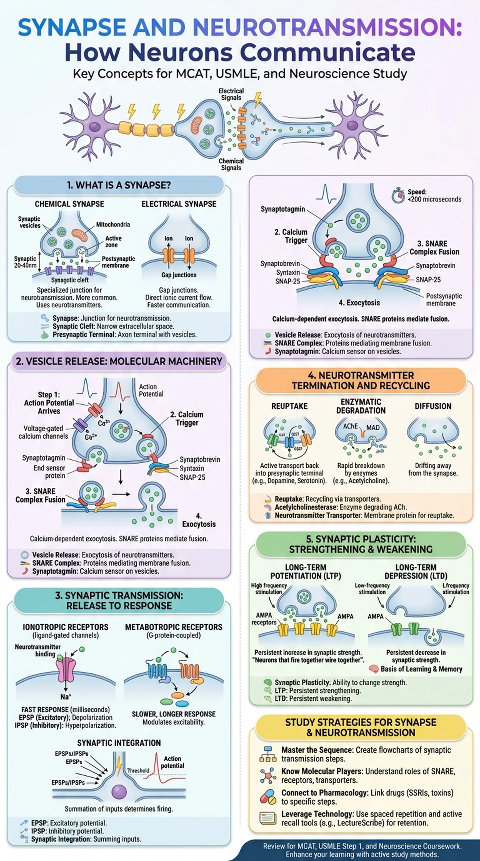
Synapses can be classified into two broad types: chemical synapses and electrical synapses. Chemical synapses, which are by far the more common type in the human nervous system, rely on the release of neurotransmitter molecules from the presynaptic terminal into the synaptic cleft, the narrow extracellular space separating the two neurons. The neurotransmitter then binds to receptors on the postsynaptic membrane to produce a response. Electrical synapses, also known as gap junctions, allow direct flow of ionic current between neurons through connexin protein channels, enabling faster but less modulable communication.

The structure of a chemical synapse includes three key components: the presynaptic terminal (also called the axon terminal or synaptic bouton), the synaptic cleft, and the postsynaptic membrane. The presynaptic terminal contains mitochondria for energy production, synaptic vesicles loaded with neurotransmitter, and an active zone where vesicle release occurs. The synaptic cleft is approximately 20 to 40 nanometers wide and contains extracellular matrix proteins that help align the presynaptic and postsynaptic specializations. Understanding synapse architecture is essential for grasping how neurotransmission translates neuronal electrical activity into the chemical signals that drive all brain function.

Key Terms

Synapse

The specialized junction between neurons where neurotransmission occurs, consisting of a presynaptic terminal, synaptic cleft, and postsynaptic membrane.

Chemical Synapse

A synapse that uses neurotransmitter molecules released into the synaptic cleft to transmit signals between neurons.

Electrical Synapse

A synapse formed by gap junctions that allows direct electrical current flow between neurons, enabling rapid communication.

Synaptic Cleft

The narrow extracellular space of approximately 20-40 nm between the presynaptic and postsynaptic membranes at a chemical synapse.

Presynaptic Terminal

The axon terminal of the transmitting neuron, containing synaptic vesicles and the molecular machinery for neurotransmitter release.

Vesicle Release: The Molecular Machinery of Neurotransmission

The process of vesicle release, also known as exocytosis, is the central event in synaptic transmission at chemical synapses. Neurotransmitters are synthesized in the neuronal cell body or axon terminal and packaged into small membrane-bound synaptic vesicles by vesicular transporters. These vesicles are then docked at the active zone of the presynaptic terminal, primed for rapid release when the appropriate signal arrives.

Vesicle release is triggered by the arrival of an action potential at the presynaptic terminal. The depolarization opens voltage-gated calcium channels, allowing calcium ions to flood into the terminal. The rise in intracellular calcium concentration is the key trigger for exocytosis. Calcium binds to synaptotagmin, a calcium sensor protein on the vesicle membrane, which then interacts with the SNARE complex to drive membrane fusion. The SNARE complex consists of three proteins: synaptobrevin (also called VAMP) on the vesicle membrane, and syntaxin and SNAP-25 on the presynaptic plasma membrane. When calcium-bound synaptotagmin engages the assembled SNARE complex, it catalyzes the fusion of the vesicle membrane with the presynaptic membrane, releasing the neurotransmitter contents into the synaptic cleft.

The speed of vesicle release is remarkable. From the moment calcium enters the terminal to the moment neurotransmitter appears in the synaptic cleft, fewer than 200 microseconds elapse. This rapidity is essential for the precise timing of neurotransmission throughout the nervous system. After fusion, the vesicle membrane is retrieved through endocytosis and recycled for subsequent rounds of vesicle release. The molecular machinery of exocytosis is a target of several biological toxins: botulinum toxin cleaves SNARE proteins to block vesicle release at the neuromuscular junction, causing paralysis, while tetanus toxin disrupts inhibitory neurotransmission in the spinal cord.

Key Terms

Vesicle Release

The calcium-dependent exocytotic process by which neurotransmitter-filled synaptic vesicles fuse with the presynaptic membrane and discharge their contents into the synaptic cleft.

SNARE Complex

A protein complex of synaptobrevin, syntaxin, and SNAP-25 that mediates the fusion of synaptic vesicles with the presynaptic membrane during neurotransmitter release.

Synaptotagmin

A calcium-sensing protein on synaptic vesicles that triggers SNARE-mediated membrane fusion upon binding calcium ions.

Voltage-Gated Calcium Channels

Ion channels in the presynaptic terminal that open in response to membrane depolarization, allowing calcium influx to trigger vesicle release.

Synaptic Transmission: From Release to Postsynaptic Response

Synaptic transmission encompasses the entire sequence of events from neurotransmitter release to the postsynaptic response. Once neurotransmitter molecules are liberated into the synaptic cleft through vesicle release, they diffuse across the narrow gap and bind to specific receptors on the postsynaptic membrane. The nature of the postsynaptic response depends on the type of receptor activated and can be either excitatory or inhibitory.

Ionotropic receptors are ligand-gated ion channels that open directly upon neurotransmitter binding, allowing rapid ion flow across the postsynaptic membrane. For example, when glutamate binds to AMPA receptors, sodium ions rush into the postsynaptic cell, producing an excitatory postsynaptic potential (EPSP) that depolarizes the membrane and brings it closer to the threshold for firing an action potential. Conversely, when GABA binds to GABA-A receptors, chloride ions enter the cell, producing an inhibitory postsynaptic potential (IPSP) that hyperpolarizes the membrane and makes firing less likely. These fast synaptic responses occur within milliseconds and are the basis of rapid point-to-point neurotransmission.

Metabotropic receptors, by contrast, are G-protein-coupled receptors that activate intracellular signaling cascades rather than directly opening ion channels. Their effects are slower in onset but longer in duration, often modulating the excitability of the neuron over seconds to minutes. The postsynaptic neuron integrates all incoming EPSPs and IPSPs through a process called synaptic integration, summing inputs in both time (temporal summation) and space (spatial summation). If the net depolarization at the axon hillock reaches threshold, the neuron fires an action potential, propagating the signal further along the neural circuit. Synaptic transmission is thus not a simple relay but a sophisticated computational process that underlies all nervous system function.

Key Terms

Synaptic Transmission

The complete process by which a chemical signal is conveyed from the presynaptic neuron across the synapse to produce a response in the postsynaptic cell.

EPSP

Excitatory postsynaptic potential; a small depolarization of the postsynaptic membrane that increases the probability of the neuron firing an action potential.

IPSP

Inhibitory postsynaptic potential; a small hyperpolarization of the postsynaptic membrane that decreases the probability of the neuron firing.

Synaptic Integration

The process by which a neuron sums all incoming excitatory and inhibitory postsynaptic potentials to determine whether to fire an action potential.

Neurotransmitter Termination and Recycling

For neurotransmission to function precisely, the signal in the synaptic cleft must be terminated rapidly after each round of synaptic transmission. If neurotransmitter molecules were allowed to persist indefinitely, receptors would be continuously activated, leading to desensitization, excitotoxicity, or loss of temporal precision. Three principal mechanisms accomplish neurotransmitter clearance from the synaptic cleft: reuptake, enzymatic degradation, and diffusion.

Reuptake is the most common termination mechanism for monoamine neurotransmitters such as dopamine, serotonin, and norepinephrine. Specific transporter proteins on the presynaptic membrane actively pump the neurotransmitter back into the presynaptic terminal, where it can be repackaged into vesicles for future vesicle release. The dopamine transporter (DAT), serotonin transporter (SERT), and norepinephrine transporter (NET) are clinically significant drug targets. Cocaine blocks DAT, increasing dopamine in the synaptic cleft and producing euphoria. SSRIs block SERT, increasing serotonin availability for treating depression.

Enzymatic degradation is the primary termination mechanism for acetylcholine at the synapse. Acetylcholinesterase (AChE), an enzyme concentrated in the synaptic cleft, rapidly hydrolyzes acetylcholine into choline and acetate. Choline is then taken back into the presynaptic terminal for resynthesis of acetylcholine. Inhibitors of AChE, such as donepezil for Alzheimer's disease and neostigmine for myasthenia gravis, prolong acetylcholine signaling by slowing its breakdown. For catecholamines, monoamine oxidase (MAO) and catechol-O-methyltransferase (COMT) degrade neurotransmitters both intracellularly and extracellularly. Diffusion, in which neurotransmitter simply drifts away from the synaptic cleft, plays a supplementary role for all transmitters. Together, these mechanisms ensure that each episode of neurotransmission is a discrete, precisely timed event within the synapse.

Key Terms

Reuptake

The process by which presynaptic transporter proteins actively remove neurotransmitter from the synaptic cleft, recycling it back into the presynaptic terminal.

Acetylcholinesterase

An enzyme in the synaptic cleft that rapidly degrades acetylcholine into choline and acetate, terminating cholinergic synaptic transmission.

Monoamine Oxidase

An intracellular enzyme that degrades monoamine neurotransmitters including dopamine, serotonin, and norepinephrine; a target of MAO inhibitor antidepressants.

Neurotransmitter Transporter

Membrane proteins such as DAT, SERT, and NET that actively transport specific neurotransmitters from the synaptic cleft back into the presynaptic neuron.

Synaptic Plasticity: How Synapses Strengthen and Weaken

Synaptic plasticity refers to the ability of synapses to strengthen or weaken over time in response to patterns of activity. This property is widely regarded as the cellular basis of learning and memory, making it one of the most important concepts in modern neuroscience. The principle was encapsulated by Donald Hebb in 1949: neurons that fire together wire together. Synaptic plasticity operates at every synapse in the nervous system and is central to understanding how neurotransmission adapts to experience.

Long-term potentiation (LTP) is the best-characterized form of synaptic strengthening. First described in the hippocampus, LTP occurs when high-frequency stimulation of a presynaptic neuron produces a persistent increase in synaptic transmission efficiency. The mechanism involves NMDA receptor activation: during strong postsynaptic depolarization, the magnesium block is relieved from the NMDA receptor channel, allowing calcium to enter the postsynaptic cell. This calcium influx activates calcium/calmodulin-dependent kinase II (CaMKII) and other signaling molecules that insert additional AMPA receptors into the postsynaptic membrane, increasing the synapse's sensitivity to future glutamate release.

Long-term depression (LTD) is the complementary process by which synaptic transmission is weakened. LTD typically results from low-frequency stimulation and involves the removal of AMPA receptors from the postsynaptic membrane through endocytosis. Both LTP and LTD can be expressed presynaptically through changes in vesicle release probability as well. The balance between potentiation and depression at individual synapses allows neural circuits to encode information, refine connections during development, and adapt to changing environmental demands. Dysfunction of synaptic plasticity mechanisms is implicated in cognitive decline associated with aging, Alzheimer's disease, and various neurodevelopmental disorders, underscoring the clinical relevance of understanding how the synapse modifies its own strength.

Key Terms

Synaptic Plasticity

The capacity of synapses to change their strength in response to activity patterns, serving as the cellular foundation of learning and memory.

Long-Term Potentiation

A persistent increase in synaptic transmission efficiency following high-frequency stimulation, mediated by NMDA receptor activation and AMPA receptor insertion.

Long-Term Depression

A persistent decrease in synaptic transmission efficiency following low-frequency stimulation, involving the removal of AMPA receptors from the postsynaptic membrane.

Hebbian Plasticity

The principle that synapses are strengthened when presynaptic and postsynaptic neurons are active simultaneously, summarized as 'neurons that fire together wire together.'

Study Strategies for Synapse and Neurotransmission

Synapse and neurotransmission topics are frequently tested on the MCAT, USMLE Step 1, and neuroscience coursework. The material involves molecular detail, sequential processes, and clinical correlations, making it well suited to structured study techniques.

First, master the sequence of synaptic transmission step by step. Create a flowchart that begins with the action potential arriving at the presynaptic terminal, proceeds through calcium influx, vesicle release via SNARE-mediated fusion, neurotransmitter diffusion across the synaptic cleft, receptor binding, postsynaptic potential generation, and finally neurotransmitter clearance. Walking through this sequence from memory is an excellent active recall exercise. Second, understand the molecular players at each stage. Know the roles of synaptotagmin and the SNARE complex in vesicle release, the distinction between ionotropic and metabotropic receptors in postsynaptic signaling, and the mechanisms of reuptake versus enzymatic degradation in signal termination.

Third, connect synapse physiology to pharmacology. Many exam questions test how drugs modify neurotransmission at specific steps. SSRIs block reuptake at the synapse, botulinum toxin prevents vesicle release, and benzodiazepines enhance postsynaptic GABA receptor function. Building a table linking each drug to its target step in synaptic transmission is an effective study tool. Fourth, learn synaptic plasticity as a story of bidirectional change. Understand why LTP and LTD require different patterns of stimulation and different downstream signaling events.

Finally, leverage technology to reinforce these concepts. Platforms like LectureScribe can transform your neuroscience lecture materials into interactive flashcards, slide presentations, and quiz questions focused on the synapse, neurotransmission, and synaptic transmission, enabling spaced repetition and active testing that strengthen long-term retention.

Key Terms

Active Recall

A study method that involves actively generating answers from memory, proven to be more effective than passive re-reading for mastering sequential processes like synaptic transmission.

Spaced Repetition

A learning technique that schedules review at progressively increasing intervals to optimize long-term memory consolidation.

MCAT

The Medical College Admission Test; requires knowledge of synapse structure, neurotransmission mechanisms, and synaptic plasticity for the biological sciences section.

Frequently Asked Questions

What is a synapse and why is it important?

A synapse is the junction between two neurons where neurotransmission occurs. It is important because it is the site where neurons communicate, enabling all brain functions from thought and movement to sensation and memory. Chemical synapses use neurotransmitters released across the synaptic cleft to transmit signals.

How does neurotransmission work step by step?

Neurotransmission begins when an action potential arrives at the presynaptic terminal, opening calcium channels. Calcium triggers vesicle release (exocytosis) of neurotransmitter into the synaptic cleft. The neurotransmitter binds postsynaptic receptors, generating excitatory or inhibitory potentials. The signal is terminated by reuptake, enzymatic degradation, or diffusion.

What is the synaptic cleft?

The synaptic cleft is the narrow extracellular gap of approximately 20-40 nanometers between the presynaptic and postsynaptic membranes at a chemical synapse. Neurotransmitters must diffuse across this space to bind postsynaptic receptors. It also contains enzymes like acetylcholinesterase that degrade certain neurotransmitters.

What is vesicle release and how is it triggered?

Vesicle release (exocytosis) is the process by which neurotransmitter-containing synaptic vesicles fuse with the presynaptic membrane and discharge their contents. It is triggered by calcium influx through voltage-gated channels, which activates synaptotagmin to drive SNARE-mediated membrane fusion.

What is the difference between synaptic transmission and neurotransmission?

The terms are closely related and often used interchangeably. Synaptic transmission specifically refers to signal transfer across a synapse, while neurotransmission is a broader term encompassing all chemical signaling between neurons. Both describe the process of converting electrical signals to chemical signals at synapses.

What is synaptic plasticity and why does it matter?

Synaptic plasticity is the ability of synapses to change their strength based on activity patterns. Long-term potentiation strengthens synapses, while long-term depression weakens them. This bidirectional modification is the cellular basis of learning and memory and is essential for neural circuit adaptation.

How do drugs affect neurotransmission at the synapse?

Drugs can modify neurotransmission at multiple steps: blocking vesicle release (botulinum toxin), enhancing or blocking postsynaptic receptors (benzodiazepines, antipsychotics), inhibiting reuptake transporters (SSRIs, cocaine), or blocking enzymatic degradation (cholinesterase inhibitors). Each mechanism alters signaling across the synaptic cleft.

Study Neuroscience Smarter

Upload your lecture notes and get AI-generated flashcards, quizzes, infographics, and study guides tailored to your curriculum.

Try LectureScribe Free

Create Study Materials

Transform your Neuroscience lectures into flashcards, quizzes, and visual study guides with AI.

  • AI-generated flashcards & quizzes
  • Visual infographics from notes
  • Interactive study shorts
Start Studying Free

Quick Facts

SubjectNeuroscience
Word Count2,500+
Slides5
InfographicIncluded
AuthorLectureScribe

Master Neuroscience with LectureScribe

Upload your lecture notes and get AI-powered flashcards, quizzes, infographics, and study guides in minutes.